. 2000 Aug 7;150(3):461-74.
doi: 10.1083/jcb.150.3.461.

Mutants affecting the structure of the cortical endoplasmic reticulum in Saccharomyces cerevisiae

Affiliations

Mutants affecting the structure of the cortical endoplasmic reticulum in Saccharomyces cerevisiae

W A Prinz et al. J Cell Biol. .

Abstract

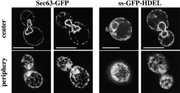
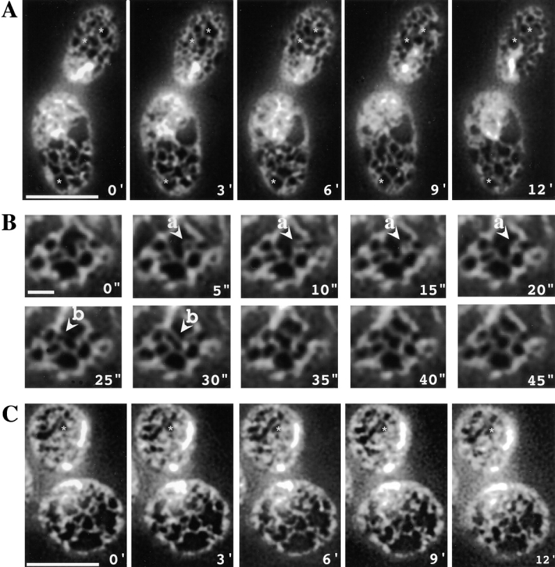
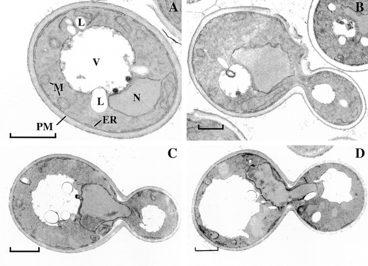
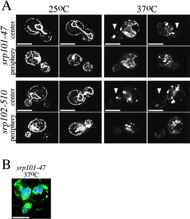
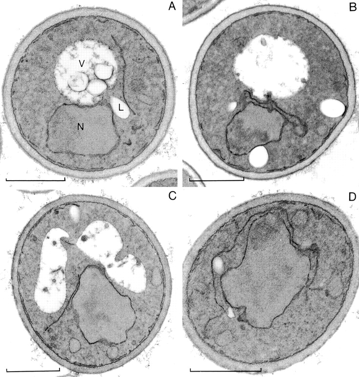
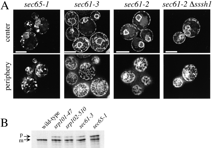
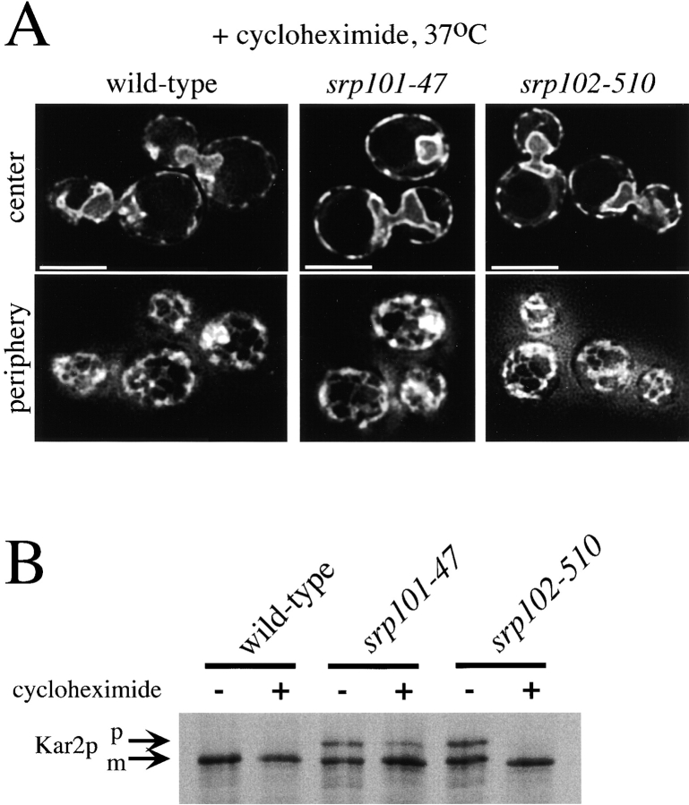
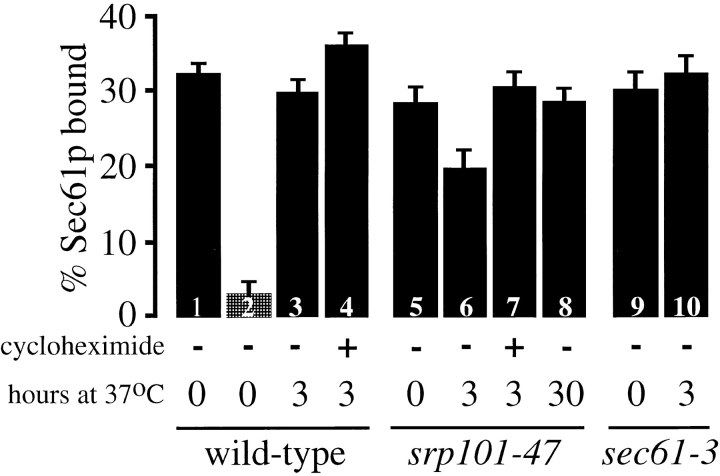
We find that the peripheral ER in Saccharomyces cerevisiae forms a dynamic network of interconnecting membrane tubules throughout the cell cycle, similar to the ER in higher eukaryotes. Maintenance of this network does not require microtubule or actin filaments, but its dynamic behavior is largely dependent on the actin cytoskeleton. We isolated three conditional mutants that disrupt peripheral ER structure. One has a mutation in a component of the COPI coat complex, which is required for vesicle budding. This mutant has a partial defect in ER segregation into daughter cells and disorganized ER in mother cells. A similar phenotype was found in other mutants with defects in vesicular trafficking between ER and Golgi complex, but not in mutants blocked at later steps in the secretory pathway. The other two mutants found in the screen have defects in the signal recognition particle (SRP) receptor. This receptor, along with SRP, targets ribosome-nascent chain complexes to the ER membrane for protein translocation. A conditional mutation in SRP also disrupts ER structure, but other mutants with translocation defects do not. We also demonstrate that, both in wild-type and mutant cells, the ER and mitochondria partially coalign, and that mutations that disrupt ER structure also affect mitochondrial structure. Our data suggest that both trafficking between the ER and Golgi complex and ribosome targeting are important for maintaining ER structure, and that proper ER structure may be required to maintain mitochondrial structure.


Figures

Figure 1
Figure 1
The peripheral ER forms a network of interconnected tubules. Fluorescence microscopy of wild-type cells expressing either Sec63-GFP or ss-GFP-HDEL. Images were acquired while focusing on either the center or periphery of the cells. Bars, 5 μm.
Figure 4
Figure 4
Mutants with conditional defects in vesicular transport between the ER and Golgi complex have altered ER structure at nonpermissive temperature. Images of cells expressing Sec63-GFP acquired while focusing on either the center or periphery of the cells. Bars, 5 μm. Arrows point to daughter cells in some cells. A, Images of sec27-95 cells grown either at 25°C or 3 h after shift to nonpermissive temperature (37°C). B, Images of sec21-1, sec23-1, and ret1-3 cells 3 h after shift to 37°C. C, Images of ufe1-1 cells 2 h after shift to 37°C and cdc48-3 cells 4 h after shift to 37°C.
Figure 2
Figure 2
Actin is required for the dynamics of the peripheral ER. Time-lapse images of cortical ER in wild-type cells expressing Sec63-GFP. A, Images of a cell taken at 3-min intervals (mother cell on bottom). Bar, 5 μm. To facilitate comparisons between frames, stars have been placed in identical places in each frame. B, Images of a portion of a cell taken at 5-s intervals. Indicated are an instance of ring closure (a) and formation of a new ER tubule (b). Bar, 1 μm. C, Images of a cell taken at 3-min intervals 10 min after 200 μM latrunculin-A was added to the medium (mother cell on bottom). Bar, 5 μm.
Figure 3
Figure 3
Neither actin filaments nor microtubules are required to maintain cortical ER structure. A, Images of the ER in wild-type cells expressing Sec63-GFP were taken after 2.5 h of growth in medium with either 20 μg/ml nocodazole or 200 μM latrunculin-A. Images were acquired while focusing on either the center or periphery of the cells. Bars, 5 μm. B, Wild-type cells expressing Sec63-GFP were fixed and stained with Alexa 594 phalloidin to visualize actin. Images were acquired while focusing on the cell cortex. Arrows point to regions in which actin filaments and ER tubules align. Bars, 5 μm.
Figure 5
Figure 5
sec27-95 cells have less peripheral ER at nonpermissive temperature. Thin-section EM of wild-type (A and B) and sec27-95 (C and D) cells grown at 25°C (A and C) or 3 h after shift to 37°C (B and D). L, Lipid droplet; M, mitochondrium; N, nucleus; PM, plasma membrane; V, vacuole; ER, endoplasmic reticulum. Bars, 1 μm.
Figure 6
Figure 6
Mutants with altered ER morphology at the nonpermissive temperature. A, Images of srp101-47 and srp102-510 cells expressing Sec63-GFP grown at either 25°C or 2 h after shift to nonpermissive temperature (37°C). Arrows point to the daughter cells in some images. Images were acquired while focusing on either the center or periphery of the cells. Bars, 5 μm. B, srp101-47 cells grown for 2 h at 37°C and stained with DAPI to visualize nuclear DNA (blue). Bar, 5 μm.
Figure 7
Figure 7
Mutants with conditional defects in SRP receptor have altered peripheral ER morphology at nonpermissive temperature. Thin-section EM of srp102-510 (A and B) and srp101-47 (C and D) cells grown at 25°C (A and C) or 3 h after shift to 37°C (B and D). L, Lipid droplet; M, mitochondrium; N, nucleus; PM, plasma membrane; V, vacuole. Bars, 1 μm.
Figure 8
Figure 8
Phenotypes of various translocation mutants. A, Images of sec65-1, sec61-3, sec61-2, and sec61-2 Δssh1 cells expressing Sec63-GFP were taken 3 h after shift to nonpermissive temperature (37°C). Images were acquired while focusing on either the center or periphery of the cells. Bars, 5 μm. B, Translocation of Kar2p was tested 1 h after shift to the nonpermissive temperature (37°C). The cells were pulse-labeled for 7 min and Kar2p was immunoprecipitated. The positions of mature Kar2p (m) and of Kar2p that has not undergone signal sequence cleavage (p) are indicated.
Figure 9
Figure 9
Cycloheximide suppresses both the ER morphology and translocation defects of SRP receptor mutants. Strains expressing Sec63-GFP were grown at 25°C and then for 6 h at 37°C with or without 1 μM cycloheximide. A, Images of the cells grown with cycloheximide. They were acquired while focusing on either the center or periphery of the cells. Bars, 5 μm. B, A portion of the cultures were pulse-labeled for 7 min and lysates were immunoprecipitated for Kar2p. m, Mature Kar2p; p, precursor of Kar2p.
Figure 10
Figure 10
Ribosome binding to Sec61p. Wild-type, sec61-3, and srp101-47 cells were grown at 25°C and then shifted to 37°C for the indicated number of hours. In some cases, cycloheximide was added to the medium when the cultures were shifted to elevated temperature. ER membranes were isolated, washed with high salt, and solubilized with digitonin. The ribosomes were pelleted and the amount of Sec61p in the pellet determined by quantitative immunoblotting. Each bar is the average of at least three independent determinations. Some samples were treated with puromycin before solubilization to release nascent polypeptides (column 2). The differences between columns 5 and 6 and columns 3 and 4 are statistically significant (P < 0.01).
Figure 11
Figure 11
The SRP receptor mutants also disrupt mitochondrial structure. Cells expressing Sec63-GFP were labeled with TMR-CH2Cl to visualize mitochondria and viewed live. Wild-type cells were grown at 30°C. The other strains were grown at 25°C and then shifted to 37°C for 2 h (srp101-47), 2.5 h (ret1-3), 4 h (mmm1-1), or 30 min (fzo1-1). Arrows point to regions of alignment of mitochondria and ER tubules in wild-type cells. Images were acquired while focusing on either the center or periphery of the cells. Bars, 5 μm.

References

    1. Achleitner G., Gaigg B., Krasser A., Kainersdorfer E., Kohlwein S.D., Perktold A., Zellnig G., Daum G. Association between the endoplasmic reticulum and mitochondria of yeast facilitates interorganelle transport of phospholipids through membrane contact. Eur. J. Biochem. 1999;264:545–553. - PubMed
    1. Amar-Costesec A., Todd J.A., Kriebich G. Segregation of the polypeptide translocation apparatus to regions of the endoplasmic reticulum containing ribophorins and ribosomes. I. functional tests on rat liver microsomal subfractions. J. Cell Biol. 1984;99:2247–2253. - PMC - PubMed
    1. Amberg D.C., Goldstein A.L., Cole C.N. Isolation and characterization of RAT1an essential gene of Saccharomyces cerevisiae required for the efficient nucleocytoplasmic trafficking of mRNA. Genes Dev. 1992;6:1173–1189. - PubMed
    1. Ayscough K.R., Stryker J., Pokala N., Sander M., Crews P., Drubin D.G. High rates of actin filament turnover in budding yeast and roles for actin in estabishment and maintenance of cell polarity revealed using the actin inhibitor latrunculin-A. J. Cell Biol. 1997;137:399–416. - PMC - PubMed
    1. Biederer T., Volkwein C., Sommer T. Degradation of subunits of the Sec61p complex, an integral component of the ER membrane, by the ubiquitin–proteasome pathway. EMBO (Eur. Mol. Biol. Organ.) J. 1996;15:2069–2076. - PMC - PubMed

Publication types

MeSH terms