. 2000 Aug 7;150(3):527-38.
doi: 10.1083/jcb.150.3.527.

Three regions within ActA promote Arp2/3 complex-mediated actin nucleation and Listeria monocytogenes motility

Affiliations

Three regions within ActA promote Arp2/3 complex-mediated actin nucleation and Listeria monocytogenes motility

J Skoble et al. J Cell Biol. .

Abstract

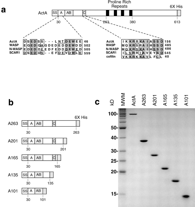
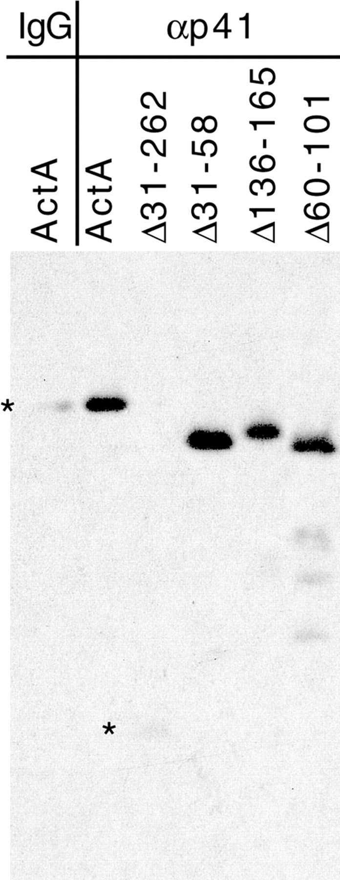
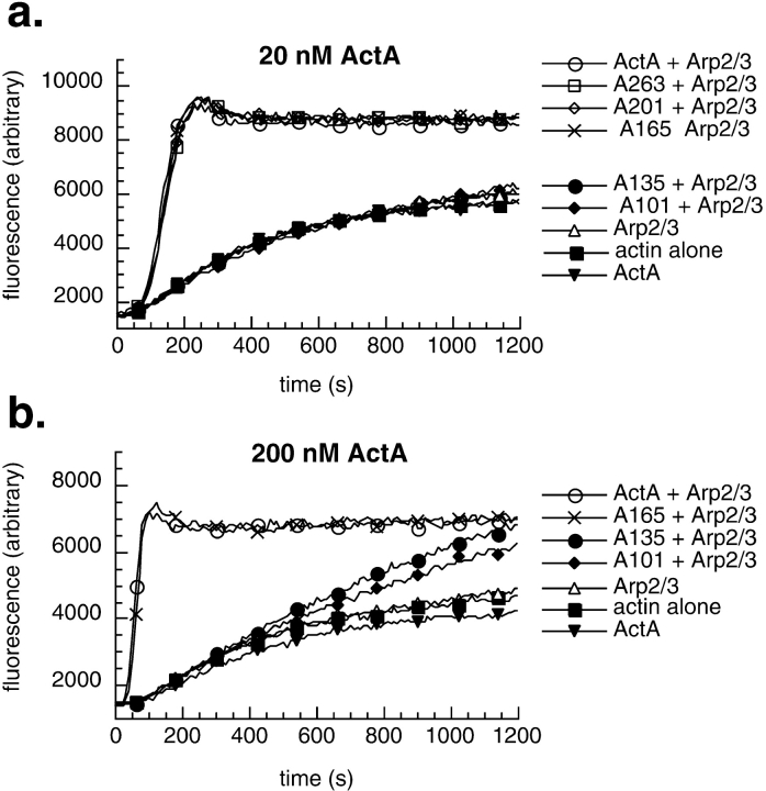
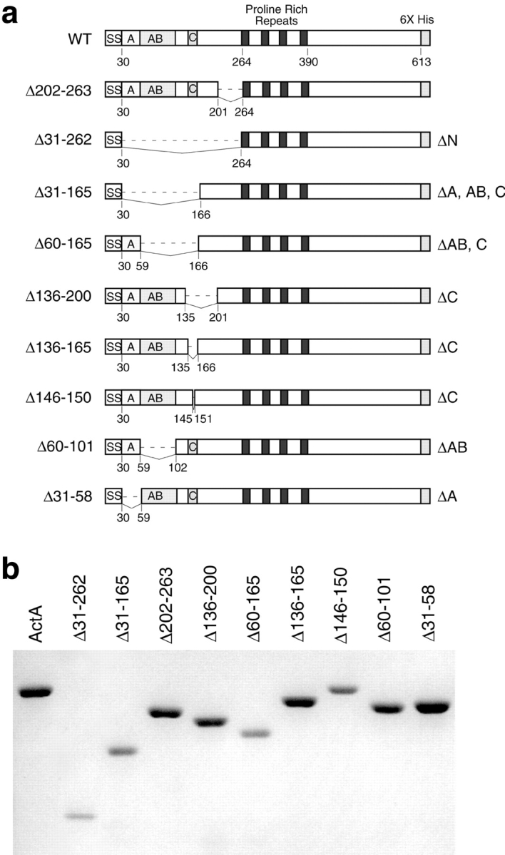
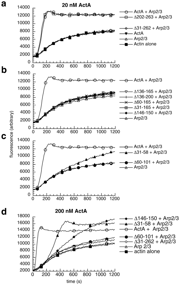
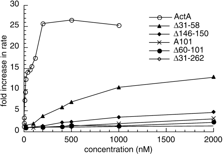
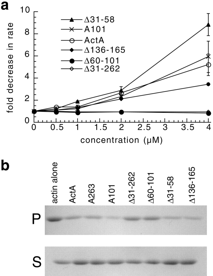
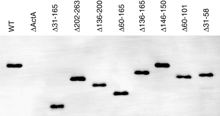
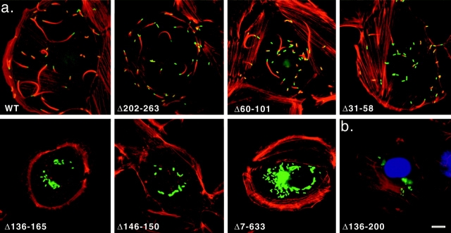
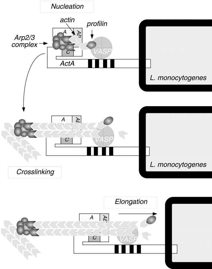
The Listeria monocytogenes ActA protein induces actin-based motility by enhancing the actin nucleating activity of the host Arp2/3 complex. Using systematic truncation analysis, we identified a 136-residue NH(2)-terminal fragment that was fully active in stimulating nucleation in vitro. Further deletion analysis demonstrated that this fragment contains three regions, which are important for nucleation and share functional and/or limited sequence similarity with host WASP family proteins: an acidic stretch, an actin monomer-binding region, and a cofilin homology sequence. To determine the contribution of each region to actin-based motility, we compared the biochemical activities of ActA derivatives with the phenotypes of corresponding mutant bacteria in cells. The acidic stretch functions to increase the efficiency of actin nucleation, the rate and frequency of motility, and the effectiveness of cell-cell spread. The monomer-binding region is required for actin nucleation in vitro, but not for actin polymerization or motility in infected cells, suggesting that redundant mechanisms may exist to recruit monomer in host cytosol. The cofilin homology sequence is critical for stimulating actin nucleation with the Arp2/3 complex in vitro, and is essential for actin polymerization and motility in cells. These data demonstrate that each region contributes to actin-based motility, and that the cofilin homology sequence plays a principal role in activation of the Arp2/3 complex, and is an essential determinant of L. monocytogenes pathogenesis.


Figures

Figure 1
Figure 1
COOH-terminal ActA truncations. (a) Schematic diagram of secreted 6xHis-tagged ActA and sequence alignments with WASP family proteins and cofilin. The signal sequence is labeled SS. Regions within the mature NH2 terminus are labeled as follows: acidic (A), actin binding (AB), and cofilin homology (C). The proline-rich repeats are shaded black, and the 6xHis tag is shaded in gray. (b) Diagram of derivatives of ActA that were truncated at amino acids 263 (A263), 201 (A201), 165 (A165), 135 (A135), and 101 (A101). (c) Purified-truncated derivatives of ActA visualized on a 15% polyacrylamide gel stained with Coomassie blue. The leftmost lane contains molecular weight markers (MWM).
Figure 7
Figure 7
Binding of ActA and ActA derivatives to Arp2/3 complex–coated resin. Equimolar concentrations of ActA or the indicated ActA derivatives were incubated with anti-p41-Arp2/3–coated resin or control IgG–coated resin. Bound ActA and ActA derivatives were resolved on a 7% polyacrylamide gel and visualized by Western blotting with polyclonal anti-ActA antibody. The expected mobility of ActA and Δ31-262 is indicated by a star.
Figure 2
Figure 2
Effects of truncated derivatives of ActA and Arp2/3 complex on actin polymerization kinetics. (a and b) Graphs of fluorescence intensity versus time after initiating actin polymerization in the pyrene-actin polymerization assay. (a) 2 μM actin in the presence or absence of 20 nM Arp2/3 complex and 20 nM ActA derivatives. (b) 2 μM actin in the presence or absence of 20 nM Arp2/3 complex and 200 nM ActA derivatives.
Figure 3
Figure 3
In-frame ActA deletions. (a) Diagram of secreted 6xHis-tagged derivatives of ActA that contain in-frame deletions within the NH2-terminal domain. Deleted residues are indicated on the left. (b) Purified derivatives of ActA containing in-frame deletions visualized on a 7.5% polyacrylamide gel stained with Coomassie blue.
Figure 4
Figure 4
Effects of ActA deletion derivatives and the Arp2/3 complex on the kinetics of actin polymerization. (a–d) Graphs of fluorescence intensity versus time after initiating polymerization in the pyrene-actin polymerization assay. (a–c) 2 μM actin in the presence or absence of 20 nM Arp2/3 complex and 20 nM ActA derivatives. (d) 2 μM actin in the presence or absence of 20 nM Arp2/3 complex and 200 nM ActA derivatives.
Figure 5
Figure 5
Graph of the fold increase in the maximal rate of pyrene-actin polymerization versus the concentration of ActA or ActA derivatives. Pyrene-actin assays were carried out in the presence of 2 μM actin, 20 nM Arp2/3 complex, and the indicated concentrations of ActA or its derivatives. The fold increase in the rate of polymerization was calculated by dividing the maximum rate of polymerization in the presence of the Arp2/3 complex and ActA or ActA derivatives by the maximum rate of polymerization in the presence of the Arp2/3 complex alone.
Figure 6
Figure 6
High concentrations of ActA and a subset of ActA derivatives slow actin polymerization in the absence of the Arp2/3 complex. (a) Graph of the fold inhibition of the maximal rate of pyrene-actin polymerization versus the concentration of ActA or ActA derivatives. The pyrene-actin assay was carried out in the presence of 2 μM actin and the indicated concentrations of ActA derivatives. The fold inhibition was calculated by dividing the maximal rate of polymerization of actin alone by the maximal rate of polymerization in the presence of ActA (bars represent SEM). (b) An actin pelleting assay was used to assess the inhibitory effect of ActA and ActA derivatives on actin polymerization. 1 μM actin was polymerized for 10 min in the presence or absence of 5 μM ActA or ActA derivatives. Actin filaments (F-actin) were separated from monomers (G-actin) by centrifugation. The amount of F-actin in the pellet (P) and G-actin in the supernatant (S) fractions was visualized on a 12% polyacrylamide gel stained with Coomassie blue.
Figure 8
Figure 8
Surface-associated ActA from actA mutants visualized by Western blotting. Surface proteins were extracted from an equivalent number of bacteria and separated on a 7% polyacrylamide gel. ActA was detected by Western blotting using polyclonal anti-ActA antibody.
Figure 9
Figure 9
Actin and L. monocytogenes visualized in infected PtK2 and HeLa cells. (a) PtK2 cells infected for 3.5 h with wild-type L. monocytogenes or the indicated mutants expressing surface-associated deletion derivatives of ActA. F-actin was visualized by staining with rhodamine-conjugated phalloidin, and bacteria were detected by indirect immunofluorescence using polyclonal anti-L. monocytogenes primary antibody followed by FITC-conjugated secondary antibody. (b) HeLa cells infected with Δ136-200 mutant L. monocytogenes for 3.5 h. F-actin was stained with rhodamine-phalloidin, DNA with DAPI, and VASP with polyclonal anti-VASP primary antibody followed by FITC-conjugated secondary antibody. Bars, 10 μm.
Figure 10
Figure 10
Model for actin nucleation by ActA and the Arp2/3 complex at the L. monocytogenes surface. The NH2-terminal domain of ActA interacts directly with the Arp2/3 complex. The acidic stretch (A) and cofilin homology sequence (C) of ActA both contribute to Arp2/3 complex activation, whereas the actin-binding region (AB) recruits and presents the actin monomer to facilitate formation of an actin nucleus. After nucleation takes place, the Arp2/3 complex dissociates from ActA, and its cross-linking activity contributes to the structure of the comet tail. During bacterial motility, profilin may speed the rate of filament elongation by delivering actin monomers to the exposed barbed ends of nucleated filaments, and VASP may bind newly formed filaments to help maintain their association with the bacterial surface.

References

    1. Banzai Y., Miki H., Yamaguchi H., Takenawa T. Essential role of neural Wiskott-Aldrich syndrome protein in neurite extension in PC12 cells and rat hippocampal primary culture cells. J. Biol. Chem. 2000;275:11987–11992. - PubMed
    1. Bachmann C., Fischer L., Walter U., Reinhard M. The EVH2 domain of the vasodilator-stimulated phosphoprotein mediates tetramerization, F-actin binding, and actin bundle formation. J. Biol. Chem. 1999;274:23549–23557. - PubMed
    1. Bi E., Zigmond S.H. Actin polymerizationwhere the WASP stings. Curr. Biol. 1999;9:R160–R163. - PubMed
    1. Brundage R.A., Smith G.A., Camilli A., Theriot J.A., Portnoy D.A. Expression and phosphorylation of the Listeria monocytogenes ActA protein in mammalian cells. Proc. Natl. Acad. Sci. 1993;USA. 90:11890–11894. - PMC - PubMed
    1. Cameron L.A., Footer M.J., van Oudenaarden A., Theriot J.A. Motility of ActA protein-coated microspheres driven by actin polymerization. Proc. Natl. Acad. Sci. USA. 1999;96:4908–4913. - PMC - PubMed

Publication types

MeSH terms