. 2000 Aug 7;150(3):581-8.
doi: 10.1083/jcb.150.3.581.

Nicotinic acid adenine dinucleotide phosphate (NAADP(+)) is an essential regulator of T-lymphocyte Ca(2+)-signaling

Affiliations

Nicotinic acid adenine dinucleotide phosphate (NAADP(+)) is an essential regulator of T-lymphocyte Ca(2+)-signaling

I Berg et al. J Cell Biol. .

Abstract

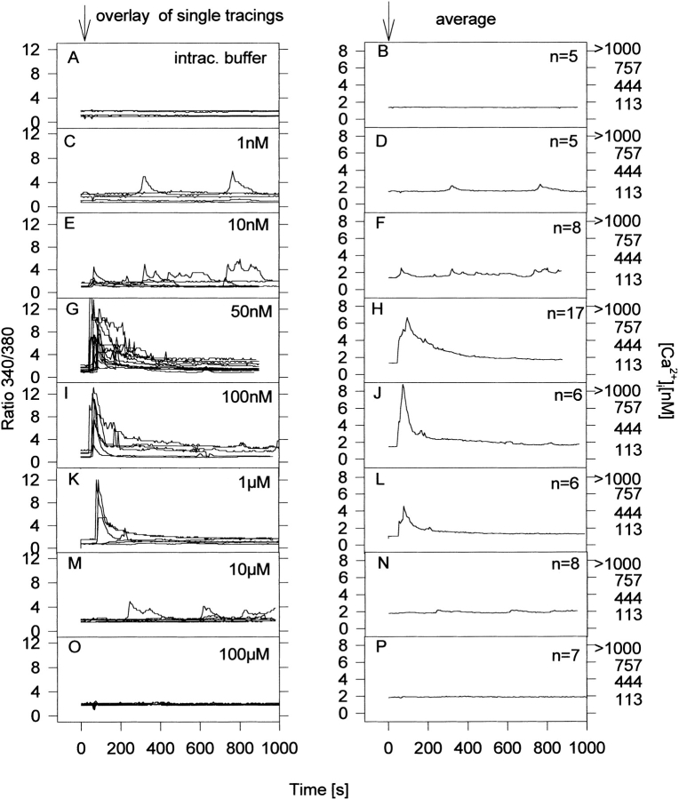
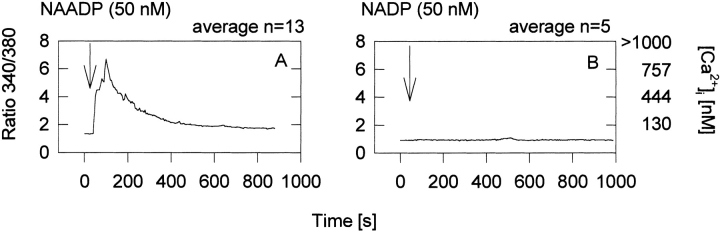
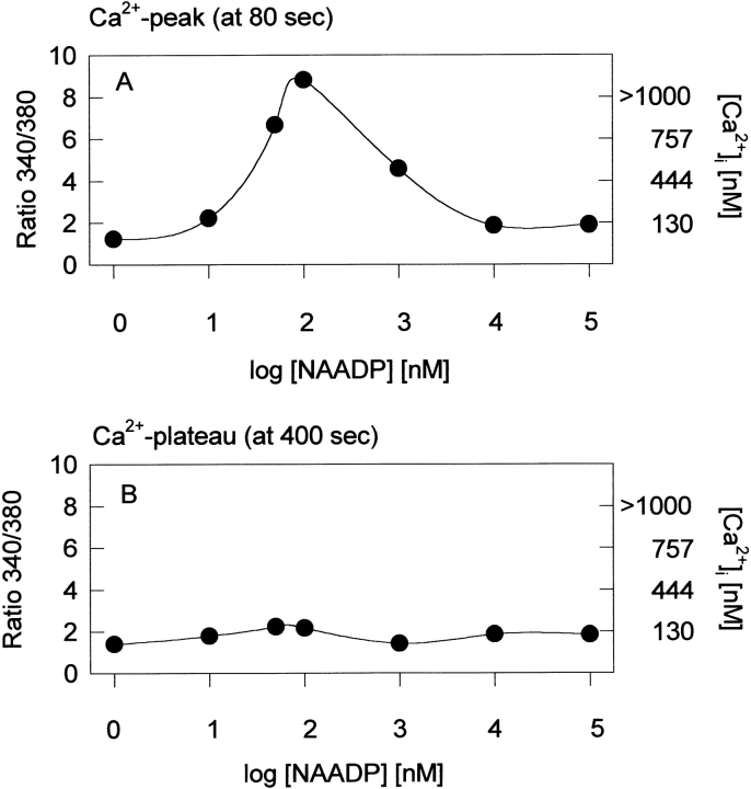
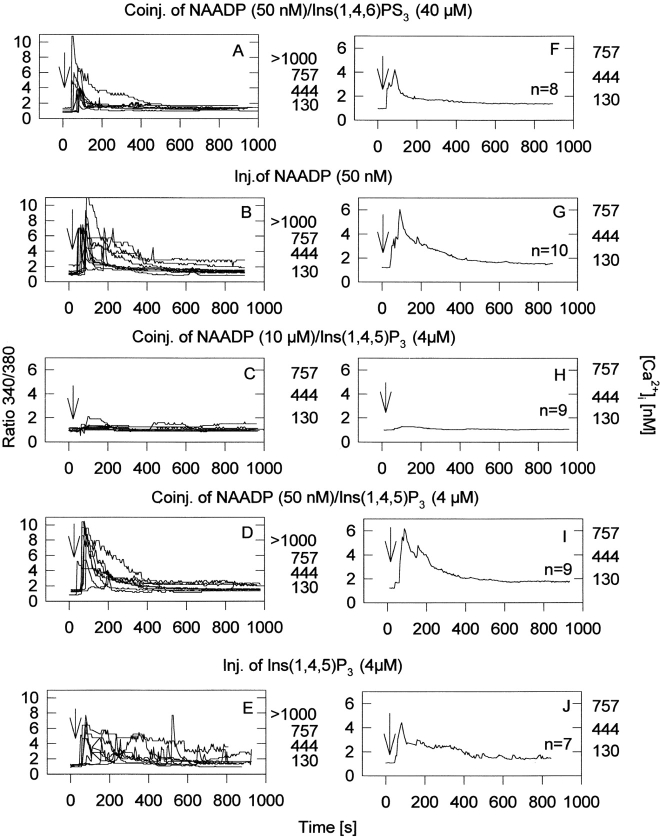
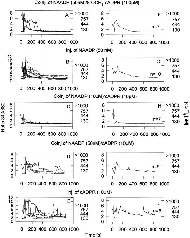
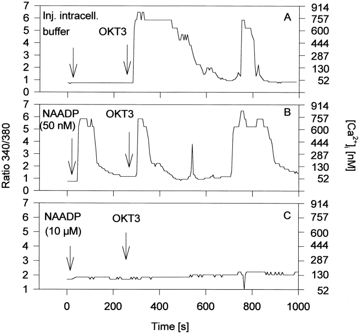
Microinjection of human Jurkat T-lymphocytes with nicotinic acid adenine dinucleotide phosphate (NAADP(+)) dose-dependently stimulated intracellular Ca(2+)-signaling. At a concentration of 10 nM NAADP(+) evoked repetitive and long-lasting Ca(2+)-oscillations of low amplitude, whereas at 50 and 100 nM, a rapid and high initial Ca(2+)-peak followed by trains of smaller Ca(2+)-oscillations was observed. Higher concentrations of NAADP(+) (1 and 10 microM) gradually reduced the initial Ca(2+)-peak, and a complete self-inactivation of Ca(2+)-signals was seen at 100 microM. The effect of NAADP(+) was specific as it was not observed with nicotinamide adenine dinucleotide phosphate. Both inositol 1,4, 5-trisphosphate- and cyclic adenosine diphosphoribose-mediated Ca(2+)-signaling were efficiently inhibited by coinjection of a self-inactivating concentration of NAADP(+). Most importantly, microinjection of a self-inactivating concentration of NAADP(+) completely abolished subsequent stimulation of Ca(2+)-signaling via the T cell receptor/CD3 complex, indicating that a functional NAADP(+) Ca(2+)-release system is essential for T-lymphocyte Ca(2+)-signaling.


Figures

Figure 1
Figure 1
Concentration-response curves of Ca2+ signals in single intact T-lymphocytes microinjected with NAADP+. Jurkat T-lymphocytes were loaded with Fura2/AM and Ca2+ was measured as detailed in Materials and Methods. Cells were injected as described in Materials and Methods in the presence of 1 mM extracellular Ca2+. Data are presented as overlays of single tracings of individual cells (left). The right shows the corresponding averages from these measurements (number of cells displayed, n = 5–17). As a control, vehicle buffer without NAADP+ was injected (A and B). The time points of microinjection are indicated by arrows.
Figure 3
Figure 3
NADP+ does not mediate Ca2+-signaling. Jurkat T-lymphocytes were loaded with Fura2/AM and ratiometric Ca2+ imaging, and parallel microinjection in the presence of 1 mM extracellular Ca2+ was carried out as detailed under Materials and Methods. Cells were injected with 50 nM NAADP+ (A) or NADP+ (B). Shown are the averages from 13 (A) and 5 (B) cells. The time points of microinjection are indicated by arrows.
Figure 2
Figure 2
Dose-response curve for NAADP+ in Jurkat T cells. Data from Fig. 1 are shown as mean values (n = 5–17) from time point 80 s (Ca2+-peak; A) or 400 s (Ca2+-plateau; B).
Figure 5
Figure 5
Influence of Ins(1,4,5)P3 and its antagonist Ins(1,4,6)PS3 on NAADP+-mediated Ca2+-signaling. Jurkat T-lymphocytes were loaded with Fura2/AM, and ratiometric Ca2+ imaging and parallel microinjection in the presence of 1 mM extracellular Ca2+ was carried out as detailed under Materials and Methods. Left, Overlays of single tracings of individual cells after injection (A–E); right, demonstrates the corresponding averages from these overlays (F–J). Shown are: A/F, coinjection of NAADP+ (50 nM) and Ins(1,4,6)PS3 (40 μM; n = 7); B/G, injection of NAADP+ (50 nM; n = 10); C/H, coinjection of NAADP+ (10 μM) and Ins(1,4,5)P3 (4 μM; n = 9); D/I, coinjection of NAADP+ (50 nM) and Ins(1,4,5)P3 (4 μM; n = 3); and E/J injection of Ins(1,4,5)P3 (4 μM; n = 8).
Figure 4
Figure 4
Influence of cADPR and its antagonist 8-OCH3-cADPR on NAADP+-mediated Ca2+-signaling. Jurkat T-lymphocytes were loaded with Fura2/AM, and ratiometric Ca2+ imaging and parallel microinjection in the presence of 1 mM extracellular Ca2+ was carried out as detailed under Materials and Methods. Left, Overlays of single tracings of individual cells after injection (A–E); right, demonstrates the corresponding averages from these overlays (F–J). Shown are (n = number of experiments): A/F, coinjection of NAADP (50 nM) and 8-OCH3-cADPR (100 μM; n = 7); B/G, injection of NAADP+ (50 nM, n = 10); C/H, coinjection of NAADP+ (10 μM) and cADPR (10μM; n = 7); D/I, coinjection of NAADP+ (50 nM) and cADPR (10 μM; n = 5); and E/J, injection of cADPR (10 μM, n = 5).
Figure 6
Figure 6
Effect of NAADP+ on OKT3-induced Ca2+-signaling in single Jurkat T-lymphocytes. Jurkat T-lymphocytes were loaded with Fura2/AM, and ratiometric Ca2+ imaging and parallel microinjection in the presence of 1 mM extracellular Ca2+ was carried out as detailed under Materials and Methods. The cells were injected with different concentrations of NAADP+ and then OKT3 (10 μg/ml) was added. Injection of intracellular buffer (A), NAADP+ (50 nM [B] and 10 μM [C]), and addition of OKT3 is indicated by arrows. Data are presented as a typical tracing from one individual cell; for each condition at least three experiments were carried out.

References

    1. Aarhus R., Dickey D.M., Graeff R.M., Gee K.R., Walseth T.F., Lee H.C. Activation and inactivation of Ca2+ release by NAADP+ . J. Biol. Chem. 1996;271:8513–8516. - PubMed
    1. Albrieux M., Lee H.C., Villaz M. Calcium signaling vy cyclic ADP-ribose, NAADP, and inositol trisphosphate are involved in distinct functions in ascidian oocytes. J. Biol. Chem. 1998;273:14566–14574. - PubMed
    1. Ashamu G.A., Galione A., Potter B.V.L. Chemo-enzymatic synthesis of analogues of the second messenger candidate cyclic adenosine 5′-diphosphate ribose. J. Chem. Soc. Chem. Commun. 1995;1359–1360
    1. Bak J., White P., Timar G., Missiaen L., Genazzani A.A., Galione A. Nicotinic acid adenine dinucleotide phosphate triggers Ca2+ release from brain microsomes. Curr. Biol. 1999;9:751–754. - PubMed
    1. Bezprozvanny I., Watras J., Ehrlich B.E. Bell-shaped calcium response curves of Ins(1,4,5)P3 and calcium-gated channels from endoplasmic reticulum of cerebellum. Nature. 1991;351:751–754. - PubMed

Publication types

MeSH terms