. 2000 Aug 15;28(16):3022-30.
doi: 10.1093/nar/28.16.3022.

Human 100-kDa homologous DNA-pairing protein is the splicing factor PSF and promotes DNA strand invasion

Affiliations

Human 100-kDa homologous DNA-pairing protein is the splicing factor PSF and promotes DNA strand invasion

A T Akhmedov et al. Nucleic Acids Res. .

Abstract

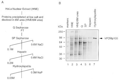
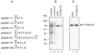
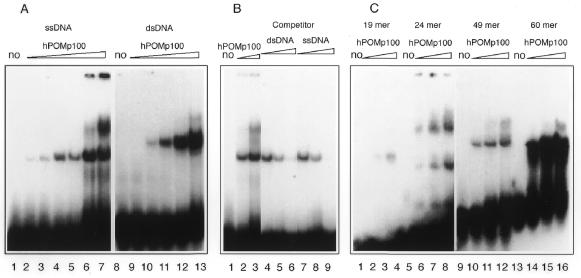
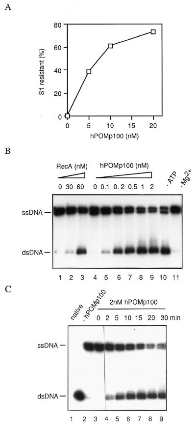
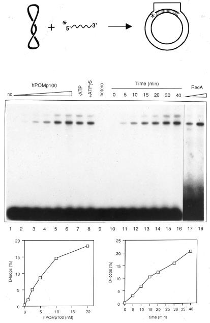
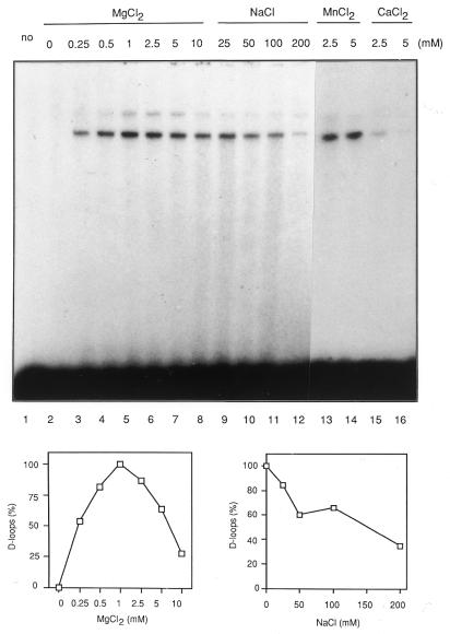
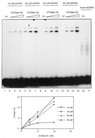
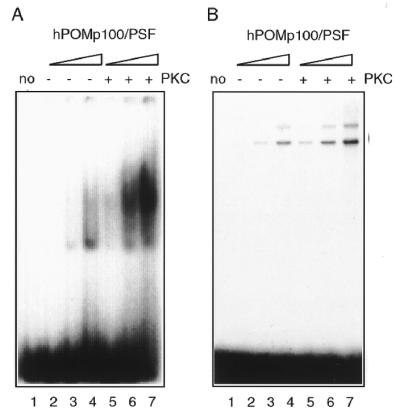
Proteins promoting homologous pairing could be involved in various fundamental biological processes. Previously we detected two mammalian nuclear proteins of 100 and 75 kDa able to promote homologous DNA pairing. Here we report isolation and characterisation of the human (h) 100-kDa DNA-pairing protein, hPOMp100, from HeLa nuclei. The peptide sequences of hPOMp100 revealed identity to the human splicing factor PSF and a DNA-binding subunit of p100/p52 heterodimer of unknown function. Bacterially expressed PSF promotes DNA pairing identical to that of hPOMp100. hPOMp100/PSF binds not only RNA but also both single-stranded (ss) and double-stranded (ds) DNA and facilitates the renaturation of complementary ssDNAs. More important, the protein promotes the incorporation of a ss oligonucleotide into a homologous superhelical dsDNA, D-loop formation. A D-loop is the first heteroduplex DNA intermediate generated between recombining DNA molecules. Moreover, this reaction could be implicated in re-establishing stalled replication forks. Consistent with this hypothesis, DNA-pairing activity of hPOMp100/PSF is associated with cellular proliferation. Significantly, phosphorylation of hPOMp100/PSF by protein kinase C inhibits its binding to RNA but stimulates its binding to DNA and D-loop formation and may represent a regulatory mechanism to direct this multifunctional protein to DNA metabolic pathways.


Figures

Figure 1
Figure 1
Purification of hPOMp100 from HeLa cell nuclei. (A) A purification scheme. (B) Coomassie-stained SDS–10% polyacrylamide gel with peak fractions from each step in (A). Lane 1, SeeBlue prestained molecular mass standards; lane 2, HNE (Fraction I); lane 3, nuclear proteins precipitated at low salt and resuspended with 6 M urea (Fraction II); lanes 4–7, fractions eluted from HiTrap Q (Fraction III), HiTrap SP (Fraction IV), HiTrap heparin (Fraction V) and Bio-Scale hydroxylapatite (Fraction VI), respectively; lane 7 contains 0.5 µg of purified hPOMp100 (Fraction VI).
Figure 2
Figure 2
Identification of hPOMp100 as the human splicing factor PSF. (A) Peptide sequences of hPOMp100. Fifty pmol of the gel-purified hPOMp100 was microsequenced and found to be the hPSF. Amino acid residues in one-letter code are numbered according to the published hPSF sequence (37). Cys431 in the reported hPSF sequence was miscalled as a Tyr (indicated in lower case) in peptide 22. It is rather unlikely that the original hPSF DNA sequencing was in error. (B) Purification of recombinant PSF (rPSF) E.coli cellular proteins after and before induction with IPTG (lanes 1 and 2, respectively); rPSF purified from E.coli and hPOMp100 purified from HeLa nuclei (Fraction VI) (lanes 3 and 4, respectively). Samples were analysed by SDS–PAGE and Coomassie staining. (C) rPSF displays the POM activity. Bacterially expressed and purified rPSF and hPOMp100 purified from HeLa nuclei (Fraction VI) were analysed by the POM assay (lanes 1 and 2, respectively). Positions of SeeBlue prestained markers and hPOMp100/PSF are indicated.
Figure 3
Figure 3
DNA-binding properties of hPOMp100/PSF. (A) Varying concentrations of hPOMp100/PSF were incubated with either 64 nM 32P-labelled 49mer oligonucleotide (lanes 1–7) or 64 nM 32P-labelled 49-bp ds-oligonucleotide (lanes 8–13). Concentrations of hPOMp100 were 0.1, 0.2, 1, 2, 10 and 20 nM (lanes 2–7) and 2, 5, 10, 20 and 40 nM (lanes 9–13). (B) 2 (lane 2) or 10 nM (lanes 3–9) hPOMp100/PSF were incubated with 64 nM 32P-labelled 49mer oligonucleotide in the absence (lanes 2 and 3) or in the presence of 25-, 50- or 100-fold molar excess of unlabelled ds- (lanes 4–6) or in the presence of 10-, 25- or 50-fold molar excess of unlabelled ssφX174 DNA (lanes 7–9). (C) 10 (lanes 2, 6, 10 and 14), 20 (lanes 3, 7, 11 and 15) or 40 nM (lanes 4, 8, 12 and 16) hPOMp100/PSF were incubated with 60 nM 32P-labelled 19 (lanes 1–4), 24 (lanes 5–8), 49 (lanes 9–12) or 60mer (lanes 13–16) oligonucleotides. The left part of the gel (lanes 1–8) was exposed 3 times longer than the right part (lanes 9–16) in order to detect a comparable amount of protein–DNA complexes. Protein–DNA complexes were analysed by native 6% PAGE. no, no protein was added.
Figure 4
Figure 4
DNA-renaturation activity of hPOMp100/PSF. (A) The S1 nuclease protection assay. The indicated amounts of hPOMp100/PSF were incubated with 1 µM heat-denatured HindIII-linearised pSV2neo [3H]DNA. The activity is expressed as percent of total DNA that is resistant to degradation with S1 nuclease. (B) The effect of hPOMp100/PSF concentration on DNA renaturation determined by the gel assay. The indicated amounts of RecA (lanes 2 and 3) or hPOMp100/PSF (lanes 5–9) were incubated with 130 nM heat-denatured 32P-labelled 422-bp DNA fragment. 2 nM hPOMp100/PSF was added in the reactions of lanes 10 and 11 and ATP or MgCl2 were omitted from the reactions in lanes 10 and 11, respectively. (C) Kinetics of DNA renaturation promoted by hPOMp100/PSF. 130 nM heat-denatured 32P-labelled 422-bp DNA fragment and 2 nM hPOMp100/PSF were incubated at 37°C for the indicated times. The products were analysed by SDS–PAGE. native, undenatured dsDNA was loaded; –hPOMp100, no protein was added, the reaction was incubated at 37°C for 30 min.
Figure 5
Figure 5
The formation of D-loops promoted by hPOMp100/PSF. Schematic diagram of DNA substrates and the expected product of the reaction is shown above the gel. Stoichiometric requirements for hPOMp100/PSF (lanes 1–6). Reactions contained 32P-labelled 49mer oligonucleotide, pUCSγ superhelical dsDNA and 1.25, 2.5, 5, 10 or 20 nM hPOMp100/PSF (lanes 2–6) were incubated at 37°C for 30 min. ATP was omitted from the reaction of lane 7 and 1 mM ATPγS was added instead of ATP in the reaction of lane 8. Heterologous M13mp18 dsDNA was added instead of pUCSγ dsDNA in the reaction of lane 9. Time course of D-loop formation (lanes 10–16). DNAs and hPOMp100/PSF (10 nM) were incubated for the indicated times. 10 nM hPOMp100/PSF was added in the reactions of lanes 7–16. RecA protein (90 or 300 nM) was added and incubated at 37°C for 30 min in the reactions of lanes 17 and 18, respectively. Reaction products were analysed by agarose gel electrophoresis as described in ‘Materials and Methods’. no, no protein was added, the reaction was incubated at 37°C for 30 min. The D-loop products were quantified and are shown below the gel.
Figure 6
Figure 6
Cofactor dependence of the hPOMp100/PSF-mediated D-loop formation. 32P-labelled 49mer oligonucleotide, pUCSγ superhelical dsDNA and 8 nM hPOMp100/PSF were incubated in reaction buffer adjusted to the indicated MgCl2 (lanes 2–8), NaCl (lanes 9–12), MnCl2 (lanes 13 and 14) or CaCl2 (lanes 15 and 16) concentrations. no, no protein was added. Reactions were incubated at 37°C for 30 min and products were analysed as described in ‘Materials and Methods’. The D-loop products were quantified and are shown below the gel.
Figure 7
Figure 7
Dependence of the hPOMp100/PSF-mediated D-loop formation on dsDNA concentration. 32P-labelled 49mer oligonucleotide and the indicated amounts of pUCSγ superhelical dsDNA were incubated with 2.5 (lanes 2, 5, 9 and 13), 5 (lanes 3, 6, 10 and 14) or 10 (lanes 4, 7, 11, 15 and 17) nM hPOMp100/PSF. 60 µM Asp700-linearised pUCSγ dsDNA was used instead of pUCSγ superhelical dsDNA in the reactions of lanes 16 and 17. no, no protein was added. Reactions were incubated at 37°C for 30 min and products were analysed as described in ‘Materials and Methods’. The D-loop products were quantified and are shown below the gel.
Figure 8
Figure 8
The PKC phosphorylation of hPOMp100/PSF stimulates its DNA binding and D-loop formation activity. Purified protein was phosphorylated in vitro by PKC as described in ‘Materials and Methods’. (A) ssDNA binding. 50 nM 32P-labelled 49mer oligonucleotide was incubated with 2.5, 5 or 10 nM hPOMp100/PSF before (lanes 2–4) or after (lanes 5–7) phosphorylation by PKC. Protein–DNA complexes were analysed by a native 6% PAGE. (B) D-loop formation. The 32P-labelled 49mer oligonucleotide and superhelical pUCSγ dsDNA were incubated with 5, 10 or 20 nM hPOMp100/PSF before (lanes 2–4) or after (lanes 5–7) phosphorylation by PKC. no, no protein was added. Reactions were incubated at 37°C for 30 min and products were analysed as described in ‘Materials and Methods’.

References

    1. Roca A.I. and Cox,M.M. (1997) Prog. Nucleic Acid. Res. Mol. Biol., 56, 129–223. - PubMed
    1. Kowalczykowski S.C. and Eggleston,A.K. (1994) Annu. Rev. Biochem., 63, 991–1043. - PubMed
    1. Shinohara A. and Ogawa,T. (1995) Trends Biochem. Sci., 20, 387–391. - PubMed
    1. Kleckner N. (1996) Proc. Natl Acad. Sci. USA, 93, 8167–8174. - PMC - PubMed
    1. Roeder G.S. (1997) Genes Dev., 11, 2600–2621. - PubMed

Publication types