. 2000 Aug 15;97(17):9437-42.
doi: 10.1073/pnas.160270597.

TARP: a nuclear protein expressed in prostate and breast cancer cells derived from an alternate reading frame of the T cell receptor gamma chain locus

Affiliations

TARP: a nuclear protein expressed in prostate and breast cancer cells derived from an alternate reading frame of the T cell receptor gamma chain locus

C D Wolfgang et al. Proc Natl Acad Sci U S A. .

Abstract

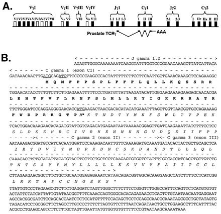
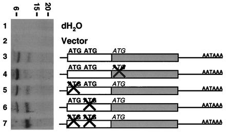
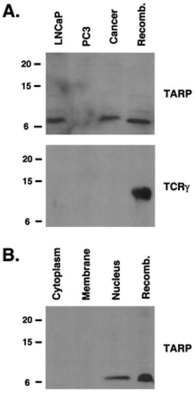
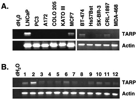
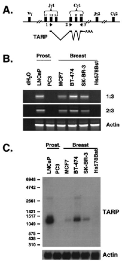
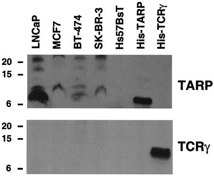
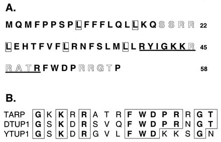
Previously, we identified the expression of a prostate-specific form of T cell receptor gamma chain (TCRgamma) mRNA in the human prostate and demonstrated that it originates from epithelial cells and not from infiltrating T lymphocytes. Here, we show that this prostate-specific transcript is also expressed in three breast cancer cell lines and breast cancer tissues. Analysis of the cDNA sequence predicts that this transcript can encode two protein products of 7 and 13 kDa, and in vitro translation experiments showed that both proteins were made. The longer ORF encodes a 13-kDa truncated version of TCRgamma, whereas the shorter alternative reading frame encodes a 7-kDa protein with five leucine residues in heptad repeats followed by a basic region. Studies with specific antibodies against each protein product revealed that both prostate and breast cancer cells contain only the 7-kDa protein, which is located in the nucleus. We have named this protein TCRgamma alternate reading frame protein (TARP). These results demonstrate that an alternative protein product is encoded by the TCRgamma locus in cells other than T lymphocytes.


Figures

Figure 1
Figure 1
The prostate-specific TCRγ transcript. (A) Schematic of the TCRγ locus and how the prostate TCRγ is transcribed and spliced in prostate cells. The transcript consists of a Jγ1.2 segment, three Cγ1 exons, and an untranslated region. (B) Nucleotide and amino acid sequences of the prostate-specific TCRγ transcript. The full-length transcript is shown starting with the transcription start site and ending with the polyadenylation signal. Arrows above the corresponding nucleotides indicate exon boundaries. The predicted amino acid sequences for two potential ORFs are noted in bold or italics. Potential initiation methionines are underlined and stop codons are outlined.
Figure 2
Figure 2
In vitro translation analysis of the prostate-specific TCRγ transcript. The prostate-specific TCRγ transcript encodes two proteins in vitro. [35S]Met-labeled in vitro-translated proteins were run on a Tris/Tricine-buffered 16.5% polyacrylamide gel and analyzed by autoradiography. A schematic representation of the mutant constructs used is shown on the right. An open box represents the first reading frame with potential initiation codons in bold, whereas the second reading frame is represented by a shaded box with the potential initiation codon in italics. “X” indicates an ATG codon mutated to ATA. Size markers in kDa are indicated on the top.
Figure 3
Figure 3
TARP is nuclear protein expressed in prostate extracts. (A) Western blot of protein extracts derived from LNCaP cells, PC3 cells, or a prostate tumor sample (Cancer). Each protein extract (20 μg) was run on a Tris/Tricine 16.5% polyacrylamide gel and probed with an antibody against TARP (Upper) or TCRγ (Lower). As a positive control, 1 μg of His-tagged TARP (Upper) or 100 ng of His-tagged TCRγ (Lower) was run on the gel (Recomb.). Size markers in kDa are indicated on the left. (B) Western blot of the cytoplasmic fraction (Cytoplasm), membrane fraction (Membrane), and nuclear fraction (Nucleus) of LNCaP cells. Each fraction (40 μg) was run on a Tris/Tricine 16.5% polyacrylamide gel and probed with an antibody against TARP. As a positive control, 1 μg of His-tagged TARP was run on the gel (Recomb.). Size markers in kDa are indicated on the left.
Figure 4
Figure 4
TARP mRNA is expressed in breast cancer cells. (A) RT-PCR was performed with primers specific for TARP (Upper) or actin (Lower) by using RNA derived from the following cell lines: prostate (LNCaP and PC3), neuroblastoma (A172), colon (COLO 205), gastric (KATO III), and breast (MCF7, BT-474, Hs57Bst, SK-BR-3, CRL-1897, and MDA-468). RT-PCRs performed without template are indicated as dH2O. (B) PCR was performed by using cDNAs derived from 12 human breast cancer tissue samples (lanes 1–12) by using primers specific for TARP (Upper) or actin (Lower). PCR reactions performed without template are indicated as dH2O. For both panels, 20% of the PCR products were run on a 1% agarose gel and visualized by ethidium bromide staining.
Figure 5
Figure 5
The TARP transcript found in the breast cell line is the same as the prostate-specific form. (A) Schematic of the TCRγ locus and how TARP is transcribed and spliced in prostate cells. Primers used for RT-PCR analysis in B are indicated. (B) RT-PCR analysis of TARP mRNA expression. PCRs with TARP primers 1 and 3 (Top), TARP primers 2 and 3 (Middle), or actin primers (Bottom) were performed with cDNAs derived from prostate cell lines (LNCaP and PC3) and breast cell lines (MCF7, BT-474, SK-BR-3, and Hs578Bst). RT-PCRs performed without template are indicated as dH2O. PCR products (20%) were run on a 1% agarose gel and visualized by ethidium bromide staining. (C) Northern blot analysis of TARP transcripts. Poly(A) mRNA (2 μg) from prostate cell lines (LNCaP and PC3) and breast cell lines (MCF7, BT-474, SK-BR-3, and Hs578Bst) were analyzed by using a constant domain fragment as probe. The autoradiograph was generated after a 24-h exposure (Upper). The same filter was stripped and analyzed with a human β-actin RNA probe to verify equal loading. The autoradiograph was generated after a 1-h exposure (Lower). RNA size markers in the nucleotides are indicated on the left.
Figure 6
Figure 6
TARP exists in the nuclei of breast cancer cells. Western blot of nuclear extracts derived from LNCaP, MCF7, BT-474, SK-BR-3, and Hs57BsT cells. Each nuclear extract (40 μg) was run on a Tris/Tricine 16.5% polyacrylamide gel and probed with an antibody against TARP (Upper) or TCRγ (Lower). As a positive control, 1 μg of His-tagged TARP (His-TARP) and 100 ng of His-tagged TCRγ (His-TCRγ) were run on the gels. Size markers in kDa are indicated on the left.
Figure 7
Figure 7
Potential functional domains of TARP. (A) TARP contains a potential leucine zipper motif and phosphorylation sites. A potential leucine zipper motif is indicated with boxed leucine followed by a basic region that is underlined. cAMP- and cGMP-dependent protein kinase phosphorylation sites (amino acids 46–49 and 55–58) and protein kinase C phosphorylation sites (amino acids 19–21 and 20–22) are outlined. (B) Protein sequence comparison of TARP with Tup1. Amino acid sequences for TARP (42–57), D. discoideum Tup1 (dTup1, 521–536), and S. cerevisiae Tup1 (yTup1, 626–660) are shown. Conserved residues are boxed.

References

    1. Hara T, Harada N, Mitsui H, Miura T, Ishizaka T, Miyajima A. Blood. 1994;84:189–199. - PubMed
    1. Liang P, Pardee A B. Science. 1992;257:967–971. - PubMed
    1. Velculescu V E, Zhang L, Vogelstein B, Kinzler K W. Science. 1995;276:1268–1272. - PubMed
    1. Schena M, Shalon D, Davis R W, Brown P O. Science. 1995;270:467–470. - PubMed
    1. Vasmatzis G, Essand M, Brinkmann U, Lee B, Pastan I. Proc Natl Acad Sci USA. 1998;95:300–304. - PMC - PubMed

MeSH terms