. 2000 Aug 15;97(17):9724-8.
doi: 10.1073/pnas.170278697.

Action potentials reliably invade axonal arbors of rat neocortical neurons

Affiliations

Action potentials reliably invade axonal arbors of rat neocortical neurons

C L Cox et al. Proc Natl Acad Sci U S A. .

Abstract

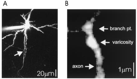
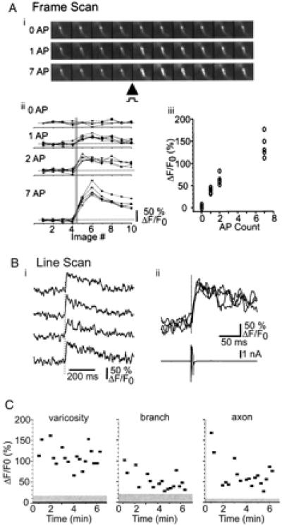
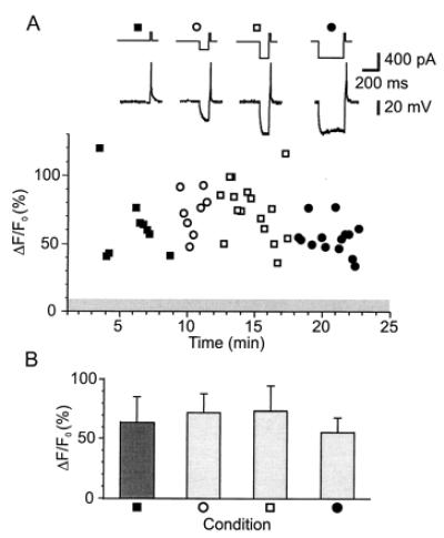
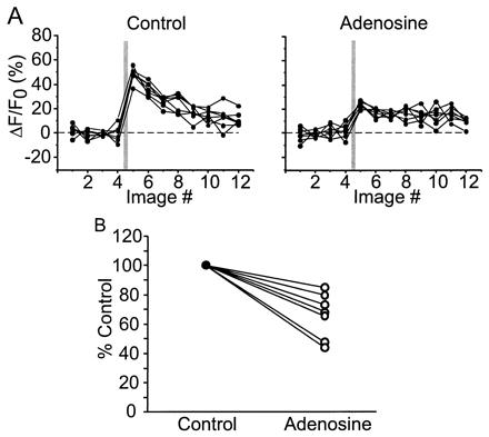
Neocortical pyramidal neurons have extensive axonal arborizations that make thousands of synapses. Action potentials can invade these arbors and cause calcium influx that is required for neurotransmitter release and excitation of postsynaptic targets. Thus, the regulation of action potential invasion in axonal branches might shape the spread of excitation in cortical neural networks. To measure the reliability and extent of action potential invasion into axonal arbors, we have used two-photon excitation laser scanning microscopy to directly image action-potential-mediated calcium influx in single varicosities of layer 2/3 pyramidal neurons in acute brain slices. Our data show that single action potentials or bursts of action potentials reliably invade axonal arbors over a range of developmental ages (postnatal 10-24 days) and temperatures (24 degrees C-30 degrees C). Hyperpolarizing current steps preceding action potential initiation, protocols that had previously been observed to produce failures of action potential propagation in cultured preparations, were ineffective in modulating the spread of action potentials in acute slices. Our data show that action potentials reliably invade the axonal arbors of neocortical pyramidal neurons. Failures in synaptic transmission must therefore originate downstream of action potential invasion. We also explored the function of modulators that inhibit presynaptic calcium influx. Consistent with previous studies, we find that adenosine reduces action-potential-mediated calcium influx in presynaptic terminals. This reduction was observed in all terminals tested, suggesting that some modulatory systems are expressed homogeneously in most terminals of the same neuron.


Figures

Figure 1
Figure 1
Imaging of axonal arbors of layer 2/3 pyramidal cells. (A) Low magnification image of a neuron filled with 100 μM OGB1, showing the apical dendrite (Top), several spiny basal dendrites, and the primary axon with two primary to secondary branch points (arrow). Note the low fluorescence intensity of secondary axonal branches compared with basal dendrites, consistent with the small diameters of axons. (B) High magnification image of a branch point between secondary and tertiary branches (same axon as in 2C). An axonal swelling is clearly recognizable by large resting fluorescence. Axonal branch points also show relatively large fluorescence.
Figure 2
Figure 2
Action-potential-evoked [Ca2+] transients in individual synaptic terminals. (A) (i) Frame scans from a single varicosity on a 2nd order axonal branch. (ii) Sequential image scans for 0, 1, 2, and 7 evoked action potentials. The solid gray line indicates timing of action potentials. (iii) Relationship between action potential number and [Ca2+] transient amplitude (three to five trials each). (B) Line scans from the same varicosity as image scans in A, showing rapid rise time of [Ca2+] transients. (i) Repeated trials illustrating [Ca2+] transients in response to single action potential. (ii) Overlay of data from i with somatic electrophysiology showing action potential in voltage clamp. (C) Amplitudes of [Ca2+] transients evoked by single action potentials in varicosities (Left), branch points (Middle), and axon (Right) (same site as in Fig. 1B). Gray regions indicate the rms noise level in trials lacking action potentials. In this example, fluorescence measurements started soon after break-in. Action-potential-evoked [Ca2+] transient amplitudes decreased with time, consistent with increasing calcium buffer concentrations.
Figure 3
Figure 3
Action potentials reliably produce [Ca2+] transients in single varicosities regardless of prior membrane potential manipulations. (A, Top) Current injection protocol used to evoke single action potential in each set of trials. (Middle) Voltage response to above protocol by somatic recordings. (Bottom) Fluorescence response amplitudes. The data plotted are peak amplitudes (ΔF/F) obtained from line scan images. In control conditions, a single depolarizing pulse evoked a single action potential, producing [Ca2+] transients (closed squares). In the next series of trials, hyperpolarizing current pulses (200 pA, 110 ms), applied to activate IA, preceded the depolarizing pulse by 2 ms. These pulses had no effect on the [Ca2+] transient amplitudes (open circles). Increasing the amplitude of the hyperpolarizing current pulse to 400 pA (open squares) and then the duration to 300 ms (closed circles) produced no significant change in the [Ca2+] transients. No failures were observed under any condition. The gray bar indicates the mean ± SD of noise measures (no stimulus). (B) The data plotted are the average responses (±SD) in each condition listed above. Essentially identical results were collected for eight other varicosities.
Figure 4
Figure 4
Adenosine suppresses [Ca2+] transients in single axonal varicosities. (A) Image scans from a single varicosity. In control conditions, clear [Ca2+] transients were observed in response to single action potentials. After bath application of adenosine (50 μM), [Ca2+] transients were significantly reduced. Each panel consists of seven consecutive trials in each condition. (B) Summary of adenosine effect in seven neurons.

Comment in

References

    1. Braitenberg V, Schutz A. Anatomy of the Cortex: Studies of Brain Function. Vol. 18. Berlin: Springer; 1991.
    1. Peters A, Palay S L, Webster HD. The Fine Structure of the Nervous System. New York: Oxford Univ. Press; 1991.
    1. Mulkey R M, Zucker R S. Nature (London) 1991;350:153–155. - PubMed
    1. Katz B. The Release of Neurotransmitter Substances. Springfield, IL: Thomas; 1969.
    1. Wall P D. Trends Neurosci. 1995;18:99–103. - PubMed

Publication types

MeSH terms

LinkOut - more resources