. 1999 Jun;1(2):123-7.
doi: 10.1038/sj.neo.7900018.

Cancer patient T cells genetically targeted to prostate-specific membrane antigen specifically lyse prostate cancer cells and release cytokines in response to prostate-specific membrane antigen

Affiliations

Cancer patient T cells genetically targeted to prostate-specific membrane antigen specifically lyse prostate cancer cells and release cytokines in response to prostate-specific membrane antigen

M C Gong et al. Neoplasia. 1999 Jun.

Abstract

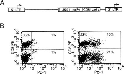
The expression of immunoglobulin-based artificial receptors in normal T lymphocytes provides a means to target lymphocytes to cell surface antigens independently of major histocompatibility complex restriction. Such artificial receptors have been previously shown to confer antigen-specific tumoricidal properties in murine T cells. We constructed a novel zeta chain fusion receptor specific for prostate-specific membrane antigen (PSMA) termed Pz-1. PSMA is a cell-surface glycoprotein expressed on prostate cancer cells and the neovascular endothelium of multiple carcinomas. We show that primary T cells harvested from five of five patients with different stages of prostate cancer and transduced with the Pz-1 receptor readily lyse prostate cancer cells. Having established a culture system using fibroblasts that express PSMA, we next show that T cells expressing the Pz-1 receptor release cytokines in response to cell-bound PSMA. Furthermore, we show that the cytokine release is greatly augmented by B7.1-mediated costimulation. Thus, our findings support the feasibility of adoptive cell therapy by using genetically engineered T cells in prostate cancer patients and suggest that both CD4+ and CD8+ T lymphocyte functions can be synergistically targeted against tumor cells.


Figures

Figure 1
Figure 1
Pz-1 vector structure and expression in primary human T cells. (A) Schematic diagram of the Pz-1 retroviral vector (Gong et al., manuscript in preparation). J591-scFv: PSMA-specific scFv derived from the monoclonal antibody (mAb) J591 [8]; CD8: CD8 hinge and transmembrane domains; zeta: T-cell receptor ζ chain cytoplasmic domain; LTR: long terminal repeat; hatched box: CD8 leader sequence; arrows indicate start of transcription. (B) FACS (Becton Dickinson & Co) analysis of transduced primary T cells. PBL harvested from cancer patients were transduced as previously described [9] and stained 3 days after retroviral infection to enumerate the fraction of Pz-1+ CD4+ and Pz-1+ CD8+ lymphocytes. Lymphocytes were incubated with an FITC-conjugated idiotype-specific antiserum, which recognizes the J591-derived scFv. After washing and incubation with 10% normal mouse serum, lymphocytes were stained with a PE-conjugated anti-CD8 mAb. Y axis: CD8PE, X axis: Pz-1 signal. Left panel: Mock transduction; right panel: Pz-1 transduction. The percentage of cells present in each quadrant is indicated.
Figure 2
Figure 2
Pz-1-transduced prostate cancer patient T lymphocytes lyse LNCaP cells. (A) LNCaP cell expression of PSMA. FACS (Becton Dickinson & Co) analysis of wild type LNCaP cells. Cells were stained with mAb J591 followed by FITC-conjugated goat anti-mouse (GAM-FITC) antibody. Y axis: cell count, X axis: fluorescence (515 nm). PSMA: PSMA signal, GAMF: GAM-FITC antibody alone signal. (B) Pz-1 directed cytotoxicity against LNCaP cells. PBL from three patients (Tables 1 and 2) were transduced with either Pz-1 or NTP [9]. Transduced PBL were used in CTL assays with LNCaP target cells. Percentage of specific lysis is represented on the Y axis, effector to target ratio on the X axis.
Figure 3
Figure 3
Pz-1-transduced prostate cancer patient T lymphocytes specifically lyse target cells expressing PSMA. (A) Left panel: FACS (Becton Dickinson & Co) analysis of wild type PC-3 cells (PSMA-) and PSMA-transduced PC-3 (PSMA+); PSMA- cells (dotted line) coincide with PC-3 cells stained with the secondary GAM-FITC antibody alone (data not shown). Cells were stained with mAb J591 [8], followed by GAM-FITC antibody. Y axis: cell count; X axis: fluorescence(515 nm). Right panel: FACS (Becton Dickinson & Co) analysis of wild type EL-4 murine thymoma cells (PSMA-) and PSMA-transduced EL-4 cells (PSMA+). (B) Pz-1-transduced PBL specifically lyse PSMA+ target cells. PBL from three patients(Tables 1 and 2) were transduced with either Pz-1 or NTP [9]. Percentage of specific lysis is represented on the Y axis, effector to target ratio on the X axis. Targets are PC3-PSMA+ and PC3-PSMA- (wild type), as described in (A, left panel). (C) Pz-1-transduced PBL specifically lyse PSMAq EL-4 cells. Pz-1q-transduced PBL of all patients effectively lysed PSMA+ targets. Background activity was variable on human target cells and uniformly low on murine target cells.
Figure 4
Figure 4
NIH3T3 fibroblast feeder cells coexpressing PSMA and B7.1 (CD80) direct cytokine release by Pz-1-transduced T cells. (A) Transduced NIH3T3 feeder cells were stained with mAb J591 as described in Figure 2 or with FITC-labeled mAb BB1 specific for B7.1. Y axis: cell count; X axis: PSMA or B7.1 signal. GAMF: GAM-FITC antibody alone. PSMA and B7.1 indicate the fluorescence obtained for each stain. (B) PSMA induced IL-2 secretion by Pz-1+ T cells. Pz-1-transduced T cells were cocultured with NIH3T3 fibroblast feeder cells. Supernatants collected 24, 48, and 72 hours after the start of the coculture were assayed for IL- 2 by ELISA. B7 + PSMA, PSMA, B7 refer to the molecules expressed by the different fibroblast monolayers. Results are from quadruplicate experiments. Error bars represent 2 standard deviations.

References

    1. Rosenberg SA, Lotze MT, Muul LM, Leitman S, Chang AE, Ettinghausen SE, Matory YL, Skibber JM, Shiloni E, Vetto JT, Seipp CA, Simpson C, Reichert CM. Observations on the systemic administration of autologous lymphokine-activated killer cells and recombinant interleukin-2 to patients with metastatic cancer. N Engl J Med. 1985;313:1485–1492. - PubMed
    1. Greenberg PD. Adoptive T cell therapy of tumors: mechanisms operative in the recognition and elimination of tumor cells. Adv Immunol. 1991;49:281–355. - PubMed
    1. Staveley-O'Carroll K, Sotomayor E, Montgomery J, Borrello I, Hwang L, Fein S, Pardoll D, Levitsky H. Induction of antigen-specific T cell anergy: An early event in the course of tumor progression. Proc Natl Acad Sci USA. 1998;95:1178–1183. - PMC - PubMed
    1. Eshhar Z, Bach N, Fitzer-Attas CJ, Gross G, Lustgarten J, Waks T, Schindler DG. The T-body approach: potential for cancer immunotherapy. Springer Semin Immunopathol. 1996;18:199–209. - PubMed
    1. Krause A, Guo H-F, Latouche J-B, Tan C, Cheung N-KV, Sadelain M. Antigen-dependent CD28 signaling selectively enhances survival and proliferation in genetically modified activated human primary T lymphocytes. J Exp Med. 1998;188:619–626. - PMC - PubMed

Publication types

MeSH terms