Review
. 2000 Jan-Apr;2(1-2):41-52.
doi: 10.1038/sj.neo.7900079.

Use of reporter genes for optical measurements of neoplastic disease in vivo

Affiliations
Review

Use of reporter genes for optical measurements of neoplastic disease in vivo

C H Contag et al. Neoplasia. 2000 Jan-Apr.

Abstract

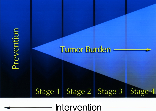
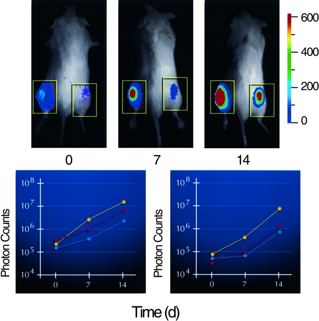
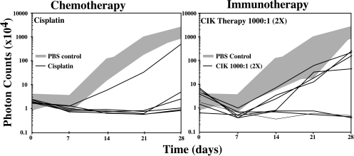
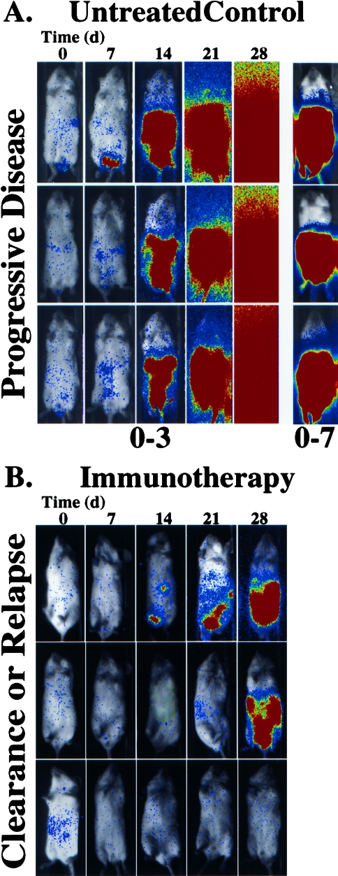
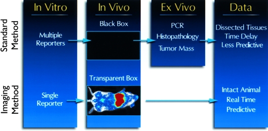
Revealing the cellular and molecular changes associated with cancer, as they occur in intact living animal models of human neoplastic disease, holds tremendous potential for understanding disease mechanisms and elucidating effective therapies. Since light is transmitted through mammalian tissues, at a low level, optical signatures conferred on tumor cells by expression of reporter genes encoding bioluminescent and fluorescent proteins can be detected externally using sensitive photon detection systems. Expression of reporter genes, such as the bioluminescent enzyme firefly luciferase (Luc) or variants of green fluorescent protein (GFP) in transformed cells, can effectively be used to reveal molecular and cellular features of neoplasia in vivo. Tumor cell growth and regression in response to various therapies have been evaluated non-invasively in living experimental animals using these reporter genes. Detection of Luc-labeled cells in vivo was extremely sensitive with signals over background from as few as 1000 human tumor cells distributed throughout the peritoneal cavity of a mouse with linear relationships between cell number and signal intensity over five logs. GFP offers the strength of high-resolution ex vivo analyses following in vivo localization of the tumor. The dynamic range of Luc detection allows the full disease course to be monitored since disease progression from small numbers of cells to extensive disease can be assessed. As such, therapies that target minimal disease as well as those designed for late stage disease can be readily evaluated in animal models. Real time spatiotemporal analyses of tumor cell growth can reveal the dynamics of neoplastic disease, and facilitate rapid optimization of effective treatment regimens. Thus, these methods improve the predictability of animal models of human disease as study groups can be followed over time, and can accelerate the development of therapeutic strategies.


Figures

Figure 1
Figure 1
New developments in cancer therapy. With the advancement of diagnostics and therapy, there is a need to develop new therapies that target small numbers of tumor cells to prevent both initiation of disease and relapse after treatment. Therefore, development of animal models that represent minimal disease is necessary to evaluate new therapeutic strategies that target these conditions.
Figure 2
Figure 2
Monitoring tumor growth at subcutaneous sites. PC-3M cells labeled with constitutive expression of a modified luciferase gene (PC-3M-luc) were injected at subcutaneous sites on each hind flank of three animals and growth of the cells was monitored by photon emission over a 14-day time course. A pseudocolor image representing light intensity is superimposed over a grayscale reference image of representative mice from each group of three (upper). Time (day) is indicated below each image. The color bar indicates average signal intensity per pixel represented by a color scheme used in the pseudocolor image. Total signal intensity over the tumor sites (boxes) was determined and plotted with respect to time for each group of three (lower). Inoculum was 1x10⁶ cells in the left flank, n=3 (left); and 1x10⁵ cells in the right flank, n=3 (right).
Figure 3
Figure 3
Effects of chemotherapy and immunotherapy on HeLa-luc cell growth in vivo. HeLa-luc cells were injected into the peritoneal cavity (time 0) at 1x10⁴ cells per animal. Groups of animals were either not treated (n=7), or treated with cis-platinum (n=6) or CIK cells at an effector to target ratio of 1000:1 (n=7). Tumor cell growth, as indicated by transmission of bioluminescent light, was assessed at weekly intervals and plotted for each animal with respect to time. The range of tumor cell growth in the untreated control animals is represented by the gray area.
Figure 4
Figure 4
Effects of immunotherapy on HeLa-luc cell growth in vivo. SCID mice bearing human xenografts, HeLa-luc cells, were either untreated (A) or treated with CIK cells at an effector to target ratio of 1000:1 (B). The growth of the tumor was followed over a 28-day time course. The pseudocolor images represent light intensity collected with a 5-minute integration time, and are all presented at the same display range (0–3 bits) with the exception of the 28-day time point for the untreated controls which is also shown at the 0–7 bit range to reveal spatial information.
Figure 5
Figure 5
Refinement of animal models using photoprotein reporters. Standard methods of monitoring tumor cell growth utilize multiple reporters and indicators to reveal the effects of experimental therapies on tumor cell growth in correlative cell culture assays. These assays often cannot be applied to in vivo analyses and assessing tumor growth in vivo is limited to measuring tumor size at superficial sites. Thus, ex vivo assays such as PCR, histological examination and weighing tumors have been necessary to assess tumor growth. These ex vivo assays often require sacrifice of the animals at multiple time points. Imaging tumor cell growth in culture and in vivo can be performed using a single reporter gene and signals for this reporter gene can be used to direct the ex vivo assays such that times and tissues can be targeted for analyses. This approach results in animal models and preclinical data that can be more predictive of human disease states.

References

    1. Clemens MJ, Bommer UA. Translational control: the cancer connection. Int J Biochem Cell Biol. 1999;31:1–23. - PubMed
    1. Dang CV, Semenza GL. Oncogenic alterations of metabolism. Trend Biochem Sci. 1999;24:68–72. - PubMed
    1. Devereux TR, Risinger JI, Barrett JC. Mutations and altered expression of the human cancer genes: what they tell us about causes. IARC Sci Publ. 1999:19–42. - PubMed
    1. Holt SE, Shay JW. Role of telomerase in cellular proliferation and cancer. J Cell Physiol. 1999;180:10–18. - PubMed
    1. Jacobson S, Pillus L. Modifying chromatin and concepts of cancer. Curr Opin Gen Dev. 1999;9:175–184. - PubMed

Publication types