

High resolution X-ray computed tomography: an emerging tool for small animal cancer research
- PMID: 10933069
- PMCID: PMC1531867
- DOI: 10.1038/sj.neo.7900069
High resolution X-ray computed tomography: an emerging tool for small animal cancer research
Abstract
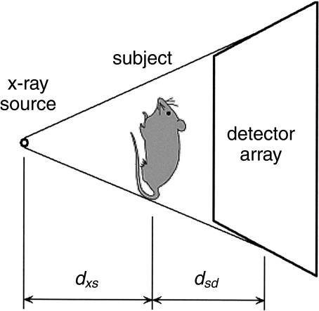
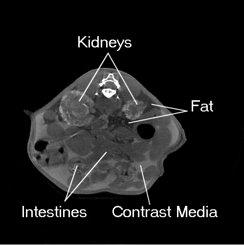
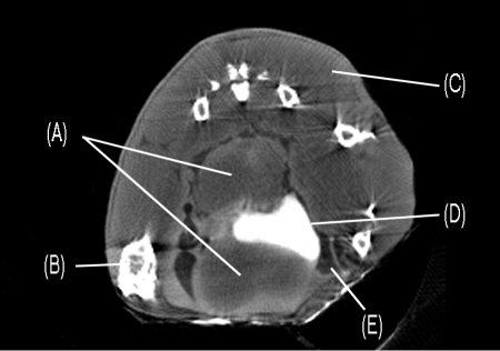
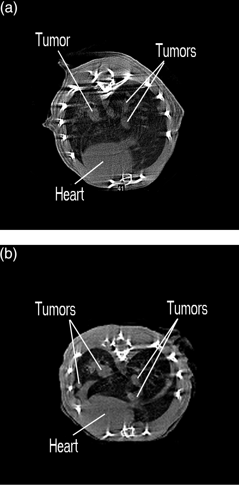
Dedicated high-resolution small animal imaging systems have recently emerged as important new tools for cancer research. These new imaging systems permit researchers to noninvasively screen animals for mutations or pathologies and to monitor disease progression and response to therapy. One imaging modality, X-ray microcomputed tomography (microCT) shows promise as a cost-effective means for detecting and characterizing soft-tissue structures, skeletal abnormalities, and tumors in live animals. MicroCT systems provide high-resolution images (typically 50 microns or less), rapid data acquisition (typically 5 to 30 minutes), excellent sensitivity to skeletal tissue and good sensitivity to soft tissue, particularly when contrast-enhancing media are employed. The development of microCT technology for small animal imaging is reviewed, and key considerations for designing small animal microCT imaging protocols are summarized. Recent studies on mouse prostate, lung and bone tumor models are overviewed.
Figures


References
-
- Hounsfield GN. Computerized transverse axial scanning (tomography): 1. Description of system. Br J Radiol. 1973;46:1016–1022. - PubMed
-
- Cromack AM. Reconstruction of densities from their projections with applications in radiological physics. Phys Med Biol. 1973;18:195–207. - PubMed
-
- Lauterbur PC. Image formation by induced local interactions — examples employing nuclear magnetic resonance. Nature. 1973;242:190–191. - PubMed
-
- Ter-Pogossian MM, Phelps ME, Mullani NA. A positron-emission transaxial tomography for nuclear imaging (PETT) Radiology. 1975;114:89–98. - PubMed
-
- Kuhl DE, Edwards RQ. Radiology. 1963;80:653–662.
Publication types
MeSH terms
LinkOut - more resources
Full Text Sources
Other Literature Sources
Medical