. 2000 Sep;74(17):8102-10.
doi: 10.1128/jvi.74.17.8102-8110.2000.

The collagen repeat sequence is a determinant of the degree of herpesvirus saimiri STP transforming activity

Affiliations

The collagen repeat sequence is a determinant of the degree of herpesvirus saimiri STP transforming activity

J K Choi et al. J Virol. 2000 Sep.

Abstract

Herpesvirus saimiri (HVS) is divided into three subgroups, A, B, and C, based on sequence divergence at the left end of genomic DNA in which the saimiri transforming protein (STP) resides. Subgroup A and C strains transform primary common marmoset lymphocytes to interleukin-2-independent growth, whereas subgroup B strains do not. To investigate the nononcogenic phenotype of the subgroup B viruses, STP genes from seven subgroup B virus isolates were cloned and sequenced. Consistent with the lack of oncogenic activity of HVS subgroup B viruses, STP-B was deficient for transforming activity in rodent fibroblast cells. Sequence comparison reveals that STP-B lacks the signal-transducing modules found in STP proteins of the other subgroups, collagen repeats and an authentic SH2 binding motif. Substitution mutations demonstrated that the lack of collagen repeats but not an SH2 binding motif contributed to the nontransforming phenotype of STP-B. Introduction of the collagen repeat sequence induced oligomerization of STP-B, resulting in activation of NF-kappaB activity and deregulation of cell growth control. These results demonstrate that the collagen repeat sequence is a determinant of the degree of HVS STP transforming activity.


Figures
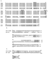
FIG. 1
FIG. 1
(A) Amino acid sequence and structural motifs of STP-B isolates. The amino acid sequences of seven STP-B clones were aligned to demonstrate similarities. TRAF-B indicates the putative TRAF-binding motif, SH2-B indicates the putative SH2-binding motif, and the grey box at the carboxyl terminus indicates the potential membrane-anchoring region. Divergent amino acid sequences are indicated with bold letters. (B) Sequence comparison of STP-A and STP-B. The grey boxes indicate the TRAF-binding motif (PxQxT/S) and SH2-binding motif (EExxYAEI/V). The bars and dots indicate identity and similarity, respectively. (C) Alignment of the TRAF-binding motifs of STP-B-SMHI with those of herpesvirus papio (HVP) LMP1. The grey boxes indicate TRAF-binding motifs.
FIG. 2
FIG. 2
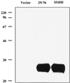
Identification of STP-B protein. 293T cells were transfected with pFJ vector, pFJ-STP-B29-76 (29-76), or pFJ-STP-B-SMHI (SMHI). Cell lysates were fractionated by SDS-PAGE, transferred to nitrocellulose, and reacted with an anti-AU-1 antibody. Sizes are shown in kilodaltons in this and subsequent figures.
FIG. 3
FIG. 3
Growth properties of Rat-1 cells expressing the STP-B gene. Puromycin-resistant cells were obtained after transfection with the retroviral vector containing STP-B-SMHI or its mutants. Puromycin-resistant cells were plated at 106 cells per 100-mm tissue culture dish. After 14 days of incubation, cells were photographed to show focus formation. Vector, Rat-babe; C488, Rat-STP-C488; SMHI, Rat-STP-B-SMHI; SMHI/EE, Rat-STP-B-SMHI/EE; SMHI/Col, Rat-STP-B-SMHI/Col; SMHI/EE/Col, Rat-STP-B-SMHI/EE/Col. Magnification, ×100.
FIG. 4
FIG. 4
Oligomerization of STP-B by collagen repeats. 293T cells were transfected with pFJ-STP-B29-76 (lane 1), pFJ-STP-B29-76/Col (lane 2), pFJ-STP-B-SMHI (lane 3), pFJ-STP-B-SMHI/EE (lane 4), pFJ-STP-B-SMHI/Col (lane 5), and pFJ (lane 6). At 48 h after transfection, heat-treated and non-heat-treated cell lysates were fractionated by SDS-PAGE, transferred to nitrocellulose, and reacted with an anti-AU-1 antibody.
FIG. 5
FIG. 5
Comparison of Src-binding activity between STP-A and STP-B. 293T cells were transfected with pFJ-Src with or without pFJ-AU1-STP-A11 (A11), pFJ-AU1-STP-B-SMHI (SMHI), and pFJ-AU1-STP-B29-76 (29-76). After 48 h, cell lysates were used for immunoprecipitation with anti-AU-1 antibody. AU-1 immune complexes were subjected to an in vitro kinase reaction. The expression level of Src, STP-A11, STP-B-SMHI, and STP-B29-76 in 293T cells was evaluated by immunoblot with anti-Src and anti-AU-1 antibodies (bottom two panels).
FIG. 6
FIG. 6
Enhanced Src-binding activity of STP-B by the substitution of negatively charged amino acids at the putative SH2-binding motif. 293T cells were transfected with pFJ-src alone (lane 2) or together with pFJ-STP-B-SMHI (lane 3), pFJ-STP-B-SMHI/EE (lane 4), pFJ-STP-B-SMHI/Col (lane 5), or pFJ-STP-B-SMHI/EE/Col (lane 6). After 48 h, cell lysates were used for immunoprecipitation (I.P.) with anti-Src 2 antibody. Src immune complexes were used for in vitro kinase assays (A). 32P-labeled proteins were separated by SDS-PAGE followed by autoradiography in a Fuji Phospho Imager. Also, Src immune complexes were separated by SDS-PAGE, transferred to nitrocellulose, and reacted with an anti-AU-1 antibody (B). The asterisk indicates the heavy chain of immunoglobulin. The same nitrocellulose membrane described above was stripped with SDS and β-mercaptoethanol and reprobed with antiphosphotyrosine (P-Y) antibody (C). The expression level of Src, STP-B-SMHI, STP-B-SMHI/EE, STP-B-SMHI/Col, and STP-B-SMHI/EE/Col in 293T cells was evaluated by immunoblot (I.B.) with anti-Src and anti-AU-1 antibodies at the bottom of panel A.
FIG. 7
FIG. 7
Interaction of STP-B with TRAFs. 293T cells were transfected with the STP-B-SMHI or STP-B29-76 expression vector together with FLAG-tagged TRAF1 or TRAF2 expression vector as shown in the figure. After 48 h, cell extracts were used for immunoprecipitations (I.P.) with an anti-FLAG antibody. The upper panels show anti-FLAG immune complexes that were subjected to immunoblot (I.B.) with an anti-AU-1 antibody to detect STP-B. The expression level of TRAFs and STP-B-SMHI or STP-B29-76 in 293T cells was evaluated by immunoblot with anti-FLAG or anti-AU-1 antibodies (bottom two panels). The asterisk indicates the heavy chain of immunoglobulin.
FIG. 8
FIG. 8
Activation of NF-κB activity by oligomerization of STP-B. Rat-1 cells stably expressing STP-B or its mutants were transfected with 5 μg of an NF-κB-driven luciferase reporter (3XkB plasmid) together with 5 μg of the pGKβgal control plasmid to measure transfection efficiency. Forty-eight hours after transfection, cell lysates were used for luciferase and β-galactosidase assays. Luciferase activity (in relative light units) was determined and normalized to β-galactosidase activities. Vector, Rat-babe; SMHI, Rat-STP-B-SMHI; SMHI/Col, Rat-STP-B-SMHI/Col; 29-76, Rat-STP-B29-76; 29-76/Col, Rat-STP-B29-76/Col. Values represent the average of two independent experiments.

Similar articles

Cited by

References

    1. Alexander L, Denekamp L, Knapp A, Auerbach M R, Damania B, Desrosiers R C. The primary sequence of rhesus monkey rhadinovirus isolate 26-95: sequence similarities to Kaposi's sarcoma-associated herpesvirus and rhesus monkey rhadinovirus isolate 17577. J Virol. 2000;74:3388–3398. - PMC - PubMed
    1. Alexander L, Du Z, Rosenzweig M, Jung J U, Desrosiers R C. A role for natural simian immunodeficiency virus and human immunodeficiency virus type 1 Nef alleles in lymphocyte activation. J Virol. 1997;71:6094–6099. - PMC - PubMed
    1. Barber R E, Kwan A P. Partial characterization of the C-terminal non-collagenous domain (NC1) of collagen type X. Biochem J. 1996;320:479–485. - PMC - PubMed
    1. Biesinger B, Müller-Fleckenstein I, Simmer B, Lang G, Wittmann S, Platzer E, Desrosiers R C, Fleckenstein B. Stable growth transformation of human T lymphocytes by herpesvirus saimiri. Proc Natl Acad Sci USA. 1992;89:3116–3119. - PMC - PubMed
    1. Biesinger B, Trimble J J, Desrosiers R C, Fleckenstein B. The divergence between two oncogenic herpesvirus saimiri strains in a genomic region related to the transforming phenotype. Virology. 1990;176:505–514. - PubMed

Publication types

MeSH terms

LinkOut - more resources