. 2000 Sep;74(17):8166-75.
doi: 10.1128/jvi.74.17.8166-8175.2000.

Regulation of the Epstein-Barr virus C promoter by AUF1 and the cyclic AMP/protein kinase A signaling pathway

Affiliations

Regulation of the Epstein-Barr virus C promoter by AUF1 and the cyclic AMP/protein kinase A signaling pathway

E M Fuentes-Pananá et al. J Virol. 2000 Sep.

Abstract

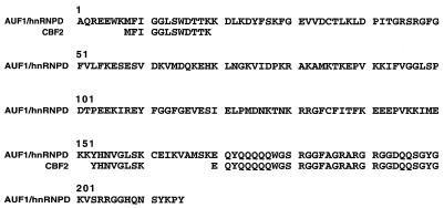
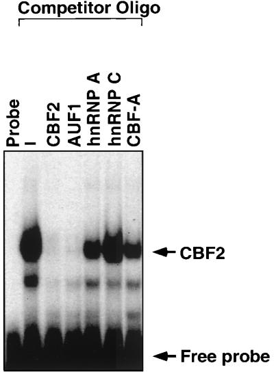
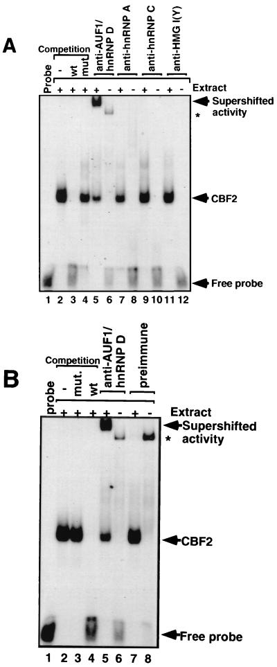
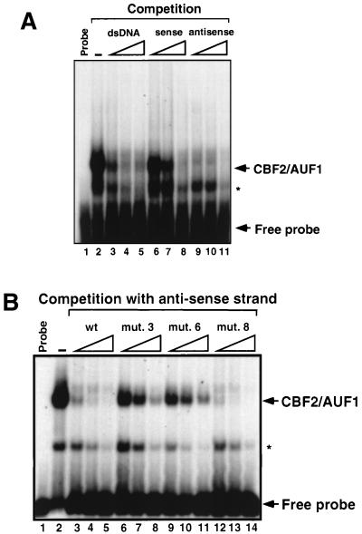
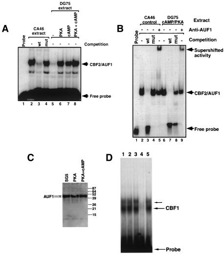
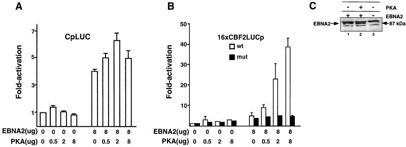
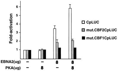
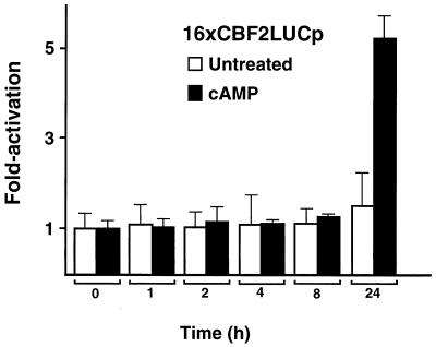
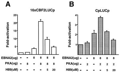
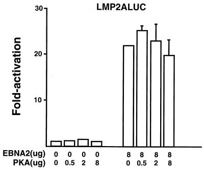
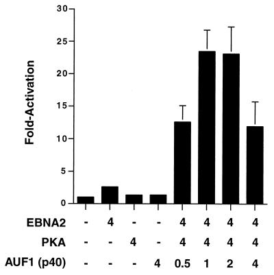
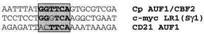
EBNA2 is an Epstein-Barr virus (EBV)-encoded protein that regulates the expression of viral and cellular genes required for EBV-driven B-cell immortalization. Elucidating the mechanisms by which EBNA2 regulates viral and cellular gene expression is necessary to understand EBV-induced B-cell immortalization and viral latency in humans. EBNA2 targets to the latency C promoter (Cp) through an interaction with the cellular DNA binding protein CBF1 (RBPJk). The EBNA2 enhancer in Cp also binds another cellular factor, C promoter binding factor 2 (CBF2), whose protein product(s) has not yet been identified. Within the EBNA2 enhancer in Cp, we have previously identified the DNA sequence required for CBF2 binding and also determined that this element is required for efficient activation of Cp by EBNA2. In this study, the CBF2 activity was biochemically purified and microsequenced. The peptides sequenced were identical to the hnRNP protein AUF1. Antibodies against AUF1 but not antibodies to related hnRNP proteins reacted with CBF2 in gel mobility shift assays. In addition, stimulation of the cellular cyclic AMP (cAMP)/protein kinase A (PKA) signal transduction pathway results in an increase in detectable CBF2/AUF1 binding activity extracted from stimulated cells. Furthermore, the CBF2 binding site was able to confer EBNA2 responsiveness to a heterologous promoter when transfected cells were treated with compounds that activate PKA or by cotransfection of plasmids expressing a constitutively active catalytic subunit of PKA. EBNA2-mediated stimulation of the latency Cp is also increased in similar cotransfection assays. These results further support an important role for CBF2 in mediating EBNA2 transactivation; they identify the hnRNP protein AUF1 as a major component of CBF2 and are also the first evidence of a cis-acting sequence other than a CBF1 binding element that is able to confer responsiveness to EBNA2.


Figures

FIG. 1
FIG. 1
Amino acid sequences of three peptides derived from purified CBF2 polypeptide aligned with the AUF1/hnRNP D protein. The sequence of the AUF1/hnRNP D protein has been reported previously (68). The peptide sequences are shown below the region matching the AUF1/hnRNP D protein sequence (top line).
FIG. 2
FIG. 2
Affinity of hnRNP binding sites for CBF2. Different hnRNP binding oligonucleotides were used to compete for CBF2 binding activity in an EMSA. CA46 nuclear extracts were mixed with a 40 M excess of the unlabeled hnRNP oligonucleotides. A 32P-labeled oligonucleotide containing the CBF2 binding site, Cp sequences −339 to −368, was then added to the shift reaction. The unlabeled competitor oligonucleotide (see Materials and Methods) used are indicated above the autoradiograph. Lanes with probe only (probe) and nuclear extract with no competitor added (−) are also shown.
FIG. 3
FIG. 3
Anti-AUF1/hnRNP D binds CBF2. Anti-hnRNP antibodies were incubated with CA46 nuclear extracts, and after 30 min of incubation, a 32P-labeled oligonucleotide probe containing the binding site for CBF2 was added to the reaction. (A) Autoradiography of a supershift analysis with antibodies against hnRNPs AUF1/hnRNP D (lane 5), A (lane 7), and C (lane 9) and control antibody against the HMG I(Y) protein (lane 11). Antibodies added to gel shift reactions without any added nuclear extract are shown in lanes 6 (AUF1/hnRNP D), 8 (hnRNP A), 10 (hnRNP C), and 12 [HMG I(Y)]. The CBF2-specific complex is confirmed by competitions with wild-type (wt) and mutant (mut) oligonucleotides shown in lanes 2 to 4. (B) Autoradiography of the supershift with preimmune and anti-AUF1/hnRNP D antisera. Lanes 1 to 6 are the same as in panel A. Preimmune serum was added to reactions in lanes 7 and 8 with and without nuclear extract, respectively. Both preimmune and immune sera form minor complexes with the labeled DNA probe and are indicated by asterisks.
FIG. 4
FIG. 4
CBF2/AUF1 is a sequence-specific DNA binding protein that binds single- and double-stranded DNA. (A) Autoradiograph of a competitive EMSA in which double- and single-stranded DNAs were used as competitors. A duplex oligonucleotide containing the Cp CBF2 binding site was used as a probe, and the same oligonucleotide in its double- or single-stranded form was used as the competitor. Competitor oligonucleotides were added at a 2.5-, 5-, and 25-fold molar excess relative to the probe, as indicated by the open triangles. Probe alone and probe bound with nuclear extract only are shown in lanes 1 and 2, respectively. (B) Autoradiograph of an EMSA in which different mutant CBF2 binding sites were used as competitors for CBF2 binding to the Cp CBF2 oligonucleotide probe. The indicated competitor oligonucleotide probes (see text) were added at a 2.5-, 5-, and 25-fold molar excess relative to the probe, as indicated by the open triangles. Probe alone and probe bound with nuclear extract only are shown in lanes 1 and 2, respectively. The asterisks denote a smaller shifted complex that is seen occasionally and is dependent on the batch of nuclear extract used for the experiments.
FIG. 5
FIG. 5
CBF2/AUF1 binding activity is increased by induction of the cAMP/PKA signal transduction pathway. (A) Autoradiograph of an EMSA that shows CBF2/AUF1 binding from heparin-Sepharose-purified CA46 control extracts (lanes 2 to 4) and cell extracts from DG75 cells that were untreated (lane 5), transfected with a plasmid constitutively expressing PKA (lane 6), treated with a cAMP analog dibutyryl cAMP (lane 7), or both (lane 8). Heparin-Sepharose-purified CA46 extracts were used as a control, and competitions with wild-type (wt) and mutant (mut) CBF2 binding oligonucleotides were used as a control for specificity of binding (lanes 2 to 4). (B) Autoradiograph of an EMSA that shows CBF2 binding from partially purified CA46 control extracts (lanes 2 to 4) and cell extracts from DG75 cells transfected with a plasmid constitutively expressing PKA and treated with the cAMP analog dibutyryl cAMP. Wild-type competitor oligonucleotide inhibits CBF2 (lanes 3 and 7), while the mutant competitor does not (lanes 4 and 8). Antibody to AUF1 was added to the gel shift reaction mixtures in lanes 5 and 9. (C) Immunoblot of AUF1 protein from SG5- or PKA-transfected DG75 cells and DG75 cells transfected with PKA and treated with dibutyryl cAMP. Blots were probed with AUF1 antiserum as described previously (66). Molecular size standards are shown on the right (in kilodaltons). AUF1 appears as a doublet of p40 and p42 proteins. (D) Autoradiograph of an EMSA that shows CBF1 binding from DG75 cells transfected with pSG5 expression vector alone (lane 1), a PKA expression vector (lane 2), or a PKA expression vector and treatment with dibutyryl cAMP (lane 3). Lanes 4 and 5 show CBF1 binding in the presence of an unlabeled wild-type CBF1 binding competitor oligonucleotide and a mutant oligonucleotide unable to bind CBF1, respectively. Probe- and CBF1-specific complexes are indicated to the right. In some cases CBF1 binding activity is detected as two bands, with the slower migrating form in less abundance (; unpublished observations).
FIG. 6
FIG. 6
Cotransfection of EBNA2 and constitutively active PKA activates Cp and confers EBNA2 responsiveness to a minimal promoter containing CBF2 binding sites. (A) Target plasmids were cotransfected with or without EBNA2- and PKA-expressing plasmids. The amounts of effector plasmids are indicated below the bar graph, and fold activation is indicated to the left. The CpLUC reporter plasmid pEFP40 was used as the target reporter plasmid. Results are averages from three independent experiments. Bars show standard errors of the means. (B) Cotransfection assays using either 16xCBF2LUC (open bars) or 16xmut.CBF2LUC (solid bars) as target plasmids and EBNA2 and/or PKA expression plasmids as effector plasmids. Concentrations of effector plasmids are indicated below the bar graph, and fold activation is indicated to the left. Target plasmids were generally used at 2.0 μg unless otherwise specified. Results are averages from three independent experiments ± standard errors of the mean. (C) Western blot to estimate the abundance of EBNA2 in transfected DG75 cells in the presence and absence of PKA induction. Extracts derived from cells transfected with FLAG-EBNA2 (lane 1), EBNA2 and PKA (lane 2), or vector alone (lane 3) were resolved by SDS-PAGE, transferred to nitrocellulose, and immunoblotted using the M5 anti-FLAG antibody. The EBNA2-specific band is indicated by the arrow on the left. Nonspecific bands were detected by the FLAG monoclonal antibody in all cellular extracts.
FIG. 7
FIG. 7
Enhancement of EBNA2-mediated stimulation of Cp is dependent on CBF1 and CBF2 binding sites. DG75 cells were transfected with target promoters CpLUC (open bars), CBF2mut.CpLUC (shaded bars), and CBF1mut.CpLUC (solid bars). Cells were harvested 48 h after transfection, and the luciferase activity was calculated. Target plasmids were cotransfected with EBNA2- or PKA-expressing plasmids alone or together. Target plasmids were generally used at 2.0 μg unless otherwise specified. Concentrations of effector plasmids used are indicated below the bar graph, and fold activation is indicated to the left. Results represent an average of three independent experiments ± standard errors of the mean.
FIG. 8
FIG. 8
Kinetics of PKA enhancement of EBNA2-mediated transactivation after treatment with dibutyryl cAMP. DG75 cells were cotransfected with the 16XCBF2LUC reporter and the EBNA2 expression plasmid (8.0 μg of each) and 2.0 μg of the 16xCBF2LUC reporter plasmid. At 24 h after transfection, cells were stimulated by adding 1 μM dibutyryl cAMP to the culture medium (solid bars). Transfected but untreated cells were used as a control (open bars). Cells were harvested at different points after induction, and the luciferase activity of the reporter gene was measured. Time points are shown below the graph in hours, and fold activation is shown on the left. Results are representative of three independent experiments ± standard errors of the mean.
FIG. 9
FIG. 9
Effect of Ser/Thr kinase inhibitor H89 on the enhancement of EBNA2-mediated stimulation of Cp by PKA. (A) DG75 cells were cotransfected with the 16xCBF2LUC reporter and EBNA2 and/or PKA expression plasmids. Target reporter plasmids were used at 2.0 μg, and the effector plasmid concentrations are indicated below the graph. The concentrations of H89 are also indicated below the graph. Cells were harvested 48 h after transfection, and the luciferase activity was calculated. Fold activation levels are shown on the left. Results represent an average of three independent experiments ± standard errors of the mean. (B) Same as panel A except the CpLUC reporter plasmid was used.
FIG. 10
FIG. 10
Analysis of the ability of EBNA2-responsive elements to be activated by PKA. DG75 cells were transfected with the LMP-2A promoter. Some cells were also transfected with EBNA2-expressing plasmids or the PKA expression plasmid. Cells were harvested 48 h after transfection, and the luciferase activity was calculated. Results represent an average of three independent experiments ± standard errors of the mean.
FIG. 11
FIG. 11
Overexpression of an AUF1 cDNA results in stimulation of a minimal promoter containing CBF2 binding sites in a dose-dependent manner. Target plasmids were cotransfected with plasmids expressing EBNA2, PKA, or AUF1 or not transfected. The amounts of effector plasmids are indicated below the bar graph, and fold activation is indicated to the left. The 16xCBF2LUC reporter plasmid was used as the target reporter plasmid. Results are averages from three independent experiments ± standard errors of the mean.
FIG. 12
FIG. 12
Comparison of the CBF2 binding sequence to AUF1 binding sequences from cellular promoters. Nucleotides shown in bold represent shared sequences from c-myc and CD21 promoters with the core CBF2 binding site. Underlined bases indicate nucleotide positions that, when mutated, affected AUF1 binding to the sequence shown.

References

    1. Armstrong R, Wen W, Meinkoth J, Taylor S, Montminy M. A refractory phase in cyclic AMP-responsive transcription requires downregulation of protein kinase A. Mol Cell Biol. 1995;15:1826–1832. - PMC - PubMed
    1. Birkenbach M, Josefsen K, Yalamanchili R, Lenoir G, Kieff E. Epstein-Barr virus-induced genes: first lymphocyte-specific G protein-coupled peptide receptors. J Virol. 1993;67:2209–2220. - PMC - PubMed
    1. Brys A, Maizels N. LR1 regulates c-myc transcription in B-cell lymphomas. Proc Natl Acad Sci USA. 1994;91:4915–4919. - PMC - PubMed
    1. Bulfone-Paus S, Dempsey L A, Maizels N. Host factors LR1 and Sp1 regulate the Fp promoter of Epstein-Barr virus. Proc Natl Acad Sci USA. 1995;92:8293–8297. - PMC - PubMed
    1. Burd C G, Dreyfuss G. RNA binding specificity of hnRNP A1: significance of hnRNP A1 high-affinity binding sites in pre-mRNA splicing. EMBO J. 1994;13:1197–1204. - PMC - PubMed

Publication types

MeSH terms

Substances

LinkOut - more resources