

doi: 10.1128/jvi.74.17.8207-8212.2000.
Transfer of specificity for human immunodeficiency virus type 1 into primary human T lymphocytes by introduction of T-cell receptor genes
Affiliations
- PMID: 10933734
- PMCID: PMC112357
- DOI: 10.1128/jvi.74.17.8207-8212.2000
Item in Clipboard
Transfer of specificity for human immunodeficiency virus type 1 into primary human T lymphocytes by introduction of T-cell receptor genes
J Virol.
2000 Sep.
Abstract
The introduction of genes encoding T-cell receptor (TCR) chains specific for human immunodeficiency virus into T cells of infected patients represents a means to quantitatively and qualitatively improve immunity to the virus. Our results demonstrate that the high level of TCR expression required for physiologic functioning can be reproducibly achieved with retroviral vectors encoding full-length unmodified TCR chains under the control of a strong internal constitutive phosphoglycerate kinase promoter.
Figures
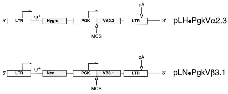
Schematic of the approximately 6-kb proviral plasmids pLH · PgkVα2.3 and pLN · PgkVβ3.1 derived from the Moloney leukemia retrovirus backbone. The Vα2.3 and Vβ3.1 TCR chains under the transcriptional control of an internal Pgk promoter were cloned into a multiple cloning site (MCS) comprised of the unique AvrII, HindIII, and ClaI (not blocked by overlapping dam methylation) sites. The plasmids contain either the hygromycin B (Hygro) or neomycin (Neo) phosphotransferase genes expressed from the 5′ LTR. The arrows indicate the direction of transcription, pA indicates the polyadenylation signal, and ψ+ indicates the packaging signal.
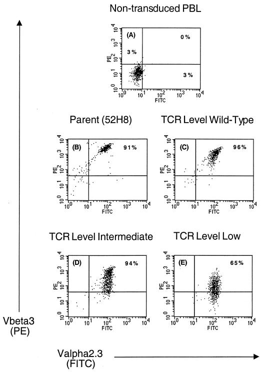
Surface expression of TCR chains specific for HIV Gag on three representative CD8+ T-cell clones transduced with the retroviruses pLH · PgkVα2.3 and pLN · PgkVβ3.1. Staining was performed with the monoclonal antibodies Vα2.3-fluorescein isothiocyanate (x axis) and biotinylated Vβ3 followed by streptavidin-phycoerythrin (y axis), and expression was assessed 21 weeks after transduction of the PBMC by flow cytometry 10 days after anti-CD3 stimulation. There was no detectable binding of isotype- and concentration-matched nonspecific antibody (data not shown). Dead cells were excluded based on uptake of propidium iodide. (A) Nontransduced autologous peripheral blood lymphocytes (PBL), used as recipient cells for introduction of the TCR genes; (B) parental clone 52H8 from which the TCR genes were isolated; (C) representative T-cell clone expressing wild-type levels of the introduced TCR chains; (D and E) Two clones expressing less-than-physiologic levels of the pairs of the introduced TCR chains.
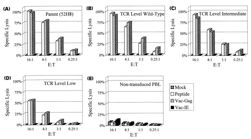
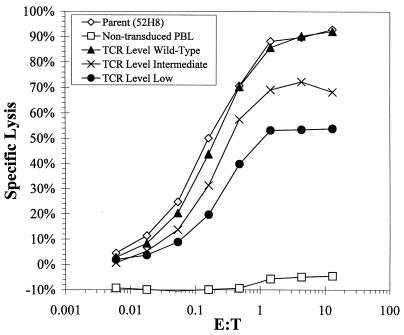
Specific lysis of HLA A3+ LCL expressing HIV Gag antigen by the parental clone 52H8 (A), transduced clones representing different levels of the introduced pair of TCR chains (B through D), and nontransduced CD8+ T cells (E). LCL targets were loaded with 5 μM concentrations of the peptide RLRPGGKKK derived from HIV Gag or were infected with recombinant vaccinia virus Gag. Control targets were mock-treated LCL and LCL infected with immediate-early vaccinia virus. Lysis was measured in a standard 5-h CRA. E:T, effector-to-target cell ratio.
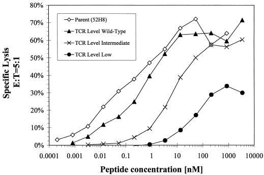
Effect of the target epitope density on lysis by the parental and transduced CTL clones. Targets were generated by incubating A3+ LCL with serial dilutions of RLRPGGKKK peptide for 5 h and lysis by clones expressing various levels of the TCR was measured in a CRA at an effector-to-target cell (E:T) ratio of 5:1.
Specific lysis of HIV-infected A3+ CD4+ Jurkat cells by CTL clones expressing various TCR levels. Jurkat cells were infected with a vesicular stomatitis virus envelope glycoprotein-pseudotyped HIV to improve the efficiency of target infection, and lysis was measured at multiple effector-to-target (E:T) ratios in a standard 5-h CRA. Lysis of noninfected mock-treated Jurkat cells was subtracted (<10% specific lysis) from lysis of HIV-infected targets. PBL, peripheral blood lymphocytes.
References
-
- Adra C N, Boer P H, McBurney M W. Cloning and expression of the mouse Pgk-1 gene and the nucleotide sequence of its promoter. Gene. 1987;60:65–74. - PubMed
-
- Altman J, Moss P A H, Goulder P, Barouch D, McHeyzer-Williams M, Bell J I, McMichael A J, Davis M M. Phenotypic analysis of antigen-specific T lymphocytes. Science. 1996;274:94–96. - PubMed
-
- Ariyoshi K, Cham F, Berry N, Jaffar S, Sabally S, Corrah T, Whittle H. HIV-2-specific cytotoxic T-lymphocyte activity is inversely related to proviral load. AIDS. 1995;9:555–559. - PubMed
-
- Barker E, Mackewicz C E, Ryes-Terán G, Sata A, Stanford S A, Fujimura S H, Christopherson C, Chang S-Y, Levy J A. Virological and immunological features of long-term human immunodeficiency virus-infected individuals who have remained asymptomatic compared with those who have progressed to acquired immunodeficiency syndrome. Blood. 1998;92:3105–3114. - PubMed
-
- Bartz S R, Vodicka M A. Production of high-titer human immunodeficiency virus type 1 pseudotyped with the vesicular stomatitis virus glycoprotein. Methods Enzymol. 1997;12:337–342. - PubMed
Publication types
MeSH terms
Substances
Grants and funding
LinkOut - more resources
Full Text Sources
Other Literature Sources
