

TPM3-ALK and TPM4-ALK oncogenes in inflammatory myofibroblastic tumors
- PMID: 10934142
- PMCID: PMC1850130
- DOI: 10.1016/S0002-9440(10)64550-6
TPM3-ALK and TPM4-ALK oncogenes in inflammatory myofibroblastic tumors
Abstract
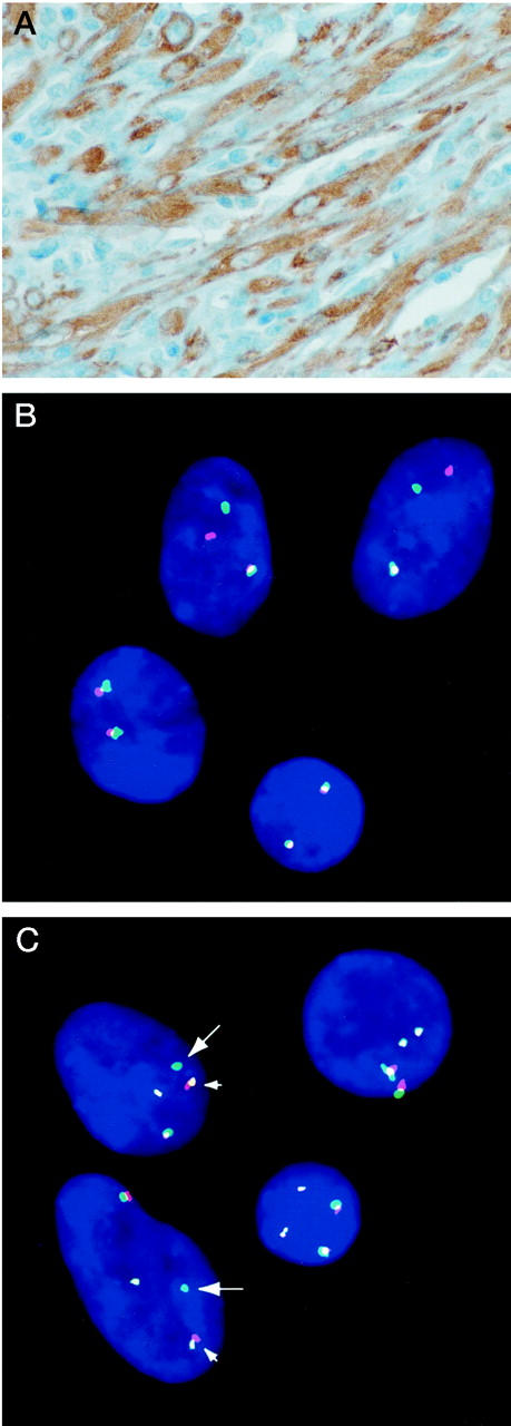
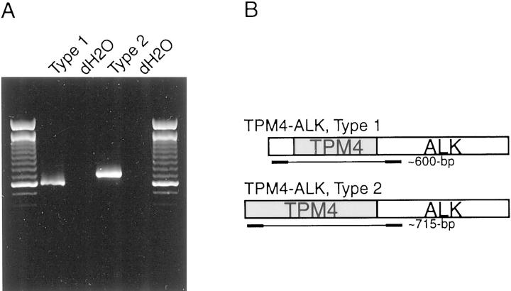
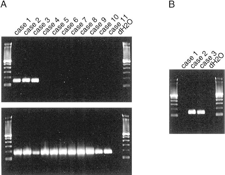
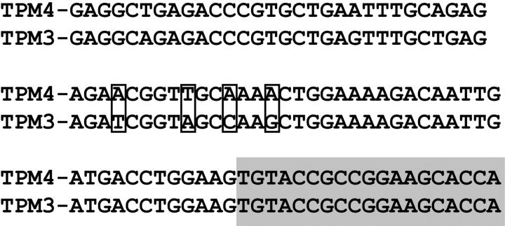
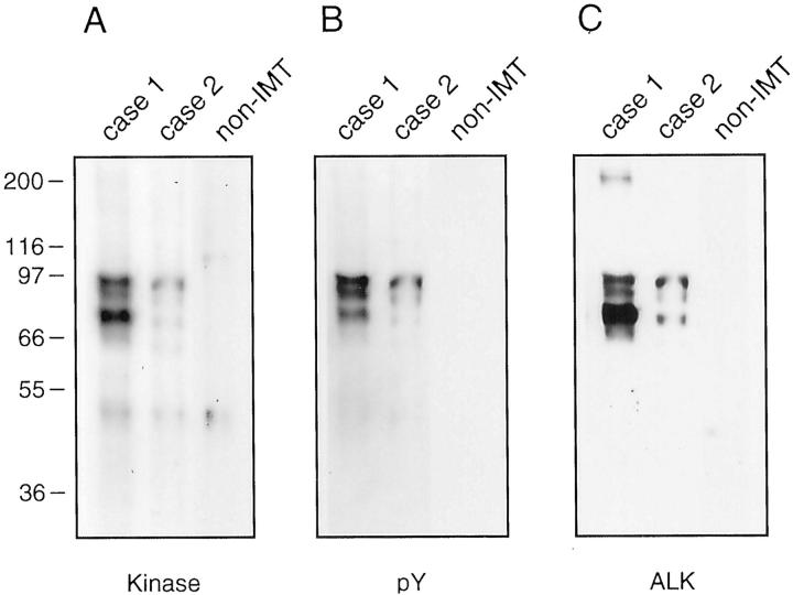
Inflammatory myofibroblastic tumors (IMTs) are neoplastic mesenchymal proliferations featuring an inflammatory infiltrate composed primarily of lymphocytes and plasma cells. The myofibroblastic cells in some IMTs contain chromosomal rearrangements involving the ALK receptor tyrosine-kinase locus region (chromosome band 2p23). ALK-which is normally restricted in its expression to neural tissues-is expressed strikingly in the IMT cells with 2p23 rearrangements. We now report a recurrent oncogenic mechanism, in IMTs, in which tropomyosin (TPM) N-terminal coiled-coil domains are fused to the ALK C-terminal kinase domain. We have cloned two ALK fusion genes, TPM4-ALK and TPM3-ALK, which encode approximately 95-kd fusion oncoproteins characterized by constitutive kinase activity and tyrosylphosphorylation. Immunohistochemical and molecular correlations, in other IMTs, implicate non-TPM ALK oncoproteins that are predominantly cytoplasmic or pre- dominantly nuclear, presumably depending on the subcellular localization of the ALK fusion partner. Notably, a TPM3-ALK oncogene was reported recently in anaplastic lymphoma, and TPM3-ALK is thereby the first known fusion oncogene that transforms, in vivo, both mesenchymal and lymphoid human cell lineages.
Figures
Comment in
-
Aberrant ALK tyrosine kinase signaling. Different cellular lineages, common oncogenic mechanisms.Am J Pathol. 2000 Aug;157(2):341-5. doi: 10.1016/S0002-9440(10)64545-2. Am J Pathol. 2000. PMID: 10934137 Free PMC article. No abstract available.
-
Sarcomatoid variant of anaplastic large cell lymphoma with cytoplasmic ALK and alpha-smooth muscle actin expression: a mimic of inflammatory myofibroblastic tumor.Am J Pathol. 2001 Jul;159(1):383-4. doi: 10.1016/s0002-9440(10)61706-3. Am J Pathol. 2001. PMID: 11438487 Free PMC article. No abstract available.
References
-
- Meis JM, Enzinger FM: Inflammatory fibrosarcoma of the mesentery and retroperitoneum. A tumor closely simulating inflammatory pseudotumor. Am J Surg Pathol 1991, 15:1146-1156 - PubMed
-
- Coffin CM, Watterson J, Priest JR, Dehner LP: Extrapulmonary inflammatory myofibroblastic tumor (inflammatory pseudotumor). A clinicopathologic and immunohistochemical study of 84 cases. Am J Surg Pathol 1995, 19:859-872 - PubMed
-
- Coffin CM, Dehner LP, Meis-Kindblom JM: Inflammatory myofibroblastic tumor, inflammatory fibrosarcoma, and related lesions: an historical review with differential diagnostic considerations. Semin Diagn Pathol 1998, 15:102-110 - PubMed
-
- Hasegawa SL, Schofield DE, Fletcher CDM: Inflammatory myofibroblastic tumor or inflammatory fibrosarcoma? Pathol Case Review 1998, 3:128-134
-
- Treissman SP, Gillis DA, Lee CL, Giacomantonio M, Resch L: Omental-mesenteric inflammatory pseudotumor. Cytogenetic demonstration of genetic changes and monoclonality in one tumor. Cancer 1994, 73:1433-1437 - PubMed
MeSH terms
Substances
Associated data
- Actions
- Actions
LinkOut - more resources
Full Text Sources
Other Literature Sources
Molecular Biology Databases
Miscellaneous
