Colorectal carcinomas arising in the hyperplastic polyposis syndrome progress through the chromosomal instability pathway
- PMID: 10934143
- PMCID: PMC1850120
- DOI: 10.1016/S0002-9440(10)64551-8
Colorectal carcinomas arising in the hyperplastic polyposis syndrome progress through the chromosomal instability pathway
Abstract
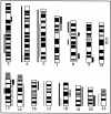
The hyperplastic polyposis syndrome is characterized by the presence within the colon of multiple large hyperplastic polyps. We describe a case of hyperplastic polyposis syndrome associated with two synchronous carcinomas, one of which arises within a pre-existing hyperplastic lesion. Comparative genomic hybridization was used to determine genetic changes in both carcinomas and several associated hyperplastic lesions. Microsatellite analysis at five loci was performed on carcinomas and representative hyperplastic polyps, and p53 status was analyzed by immunohistochemistry. Both carcinomas showed multiple genetic aberrations, including high level gains of 8q and 13q, and loss of 5q. These changes were not seen in the hyperplastic polyps. Microsatellite instability was not seen in the carcinomas, four separate hyperplastic polyps, the hyperplastic polyp with mild adenomatous change associated with the carcinoma, or a separate serrated adenoma. Allelic imbalance in the cancers at D5S346 and D17S938 suggested allelic loss of both p53 and APC, as well as at the loci D13S263, D13S174, D13S159, and D18S49. An early invasive carcinoma in one hyperplastic polyp stained for p53 protein, but the associated hyperplastic polyp was negative. In this case, neoplastic progression followed the typical genetic pathway of common colorectal carcinoma and occurred synchronously with mutation of p53.
Figures


Comment in
-
Biological significance of microsatellite instability-low (MSI-L) status in colorectal tumors.Am J Pathol. 2001 Feb;158(2):779-81. doi: 10.1016/s0002-9440(10)64020-5. Am J Pathol. 2001. PMID: 11159215 Free PMC article. No abstract available.
References
-
- Fujiwara T, Stolker JM, Watanabe T, Rashid A, Longo P, Eshleman JR, Booker S, Lynch HT, Jass JR, Green JS, Kim H, Jen J, Vogelstein B, Hamilton SR: Accumulated clonal genetic alterations in familial and sporadic colorectal carcinomas with widespread instability in microsatellite sequences. Am J Pathol 1998, 153:1063-1078 - PMC - PubMed
-
- Williams GT, Arthur JF, Bussey HJ, Morson BC: Metaplastic polyps and polyposis of the colorectum. Histopathology 1980, 4:155-170 - PubMed
-
- Jorgensen H, Mogensen AM, Svendsen LB: Hyperplastic polyposis of the large bowel. Three cases and a review of the literature. Scand J Gastroenterol 1996, 31:825-830 - PubMed
-
- Orii S, Nakamura S, Sugai T, Habano W, Akasaka I, Nakasima F, Kazama H, Hasimoto Y, Takahasi H, Sugawara M, Sato S: Hyperplastic (metaplastic) polyposis of the colorectum associated with adenomas and an adenocarcinoma. J Clin Gastroenterol 1997, 25:369-372 - PubMed
-
- McCann BG: A case of metaplastic polyposis of the colon associated with focal adenomatous change and metachronous adenocarcinomas. Histopathology 1988, 13:700-702 - PubMed
Publication types
MeSH terms
Substances
LinkOut - more resources
Full Text Sources
Medical
Research Materials
Miscellaneous

