. 2000 Aug;157(2):647-58.
doi: 10.1016/S0002-9440(10)64575-0.

Proinflammatory cytokine, chemokine, and cellular adhesion molecule expression during the acute phase of experimental brain abscess development

Affiliations

Proinflammatory cytokine, chemokine, and cellular adhesion molecule expression during the acute phase of experimental brain abscess development

T Kielian et al. Am J Pathol. 2000 Aug.

Abstract

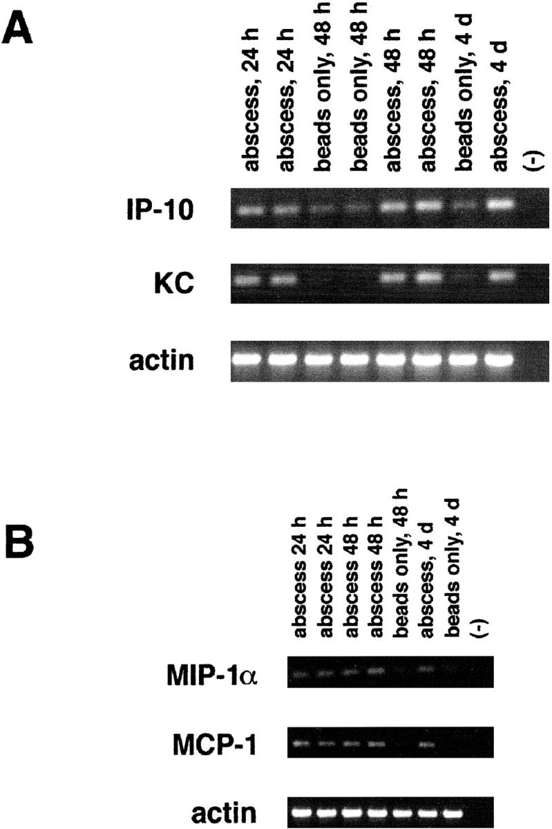
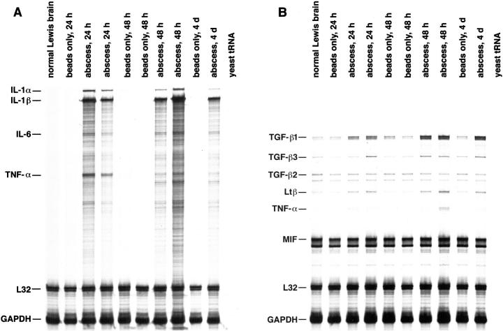
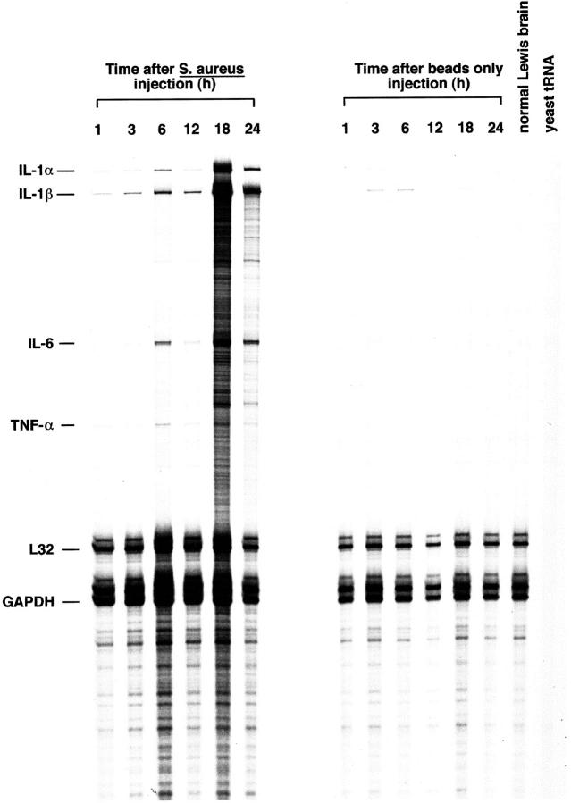
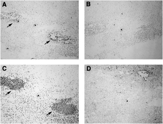
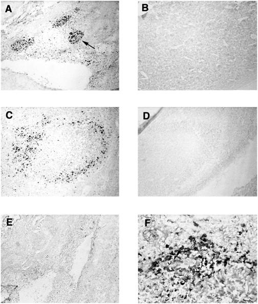
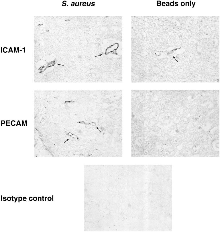
Brain abscess represents the infectious disease sequelae associated with the influx of inflammatory cells and activation of resident parenchymal cells in the central nervous system. However, the immune response leading to the establishment of a brain abscess remains poorly defined. In this study, we have characterized cytokine and chemokine expression in an experimental brain abscess model in the rat during the acute stage of abscess development. RNase protection assay revealed the induction of the proinflammatory cytokines interleukin (IL)-1alpha, IL-1beta, IL-6, and tumor necrosis factor-alpha as early as 1 to 6 hours after Staphylococcus aureus exposure. Evaluation of chemokine expression by reverse transcription-polymerase chain reaction demonstrated enhanced levels of the CXC chemokine KC 24 hours after bacterial exposure, which correlated with the appearance of neutrophils in the abscess. In addition, two CC chemokines, monocyte chemoattractant protein-1 and macrophage inflammatory protein-1alpha were induced within 24 hours after S. aureus exposure and preceded the influx of macrophages and lymphocytes into the brain. Analysis of abscess lesions by in situ hybridization identified CD11b+ cells as the source of IL-1beta in response to S. aureus. Both intercellular adhesion molecule-1 and platelet endothelial cell adhesion molecule expression were enhanced on microvessels in S. aureus but not sterile bead-implanted tissues at 24 and 48 hours after treatment. These results characterize proinflammatory cytokine and chemokine expression during the early response to S. aureus in the brain and provide the foundation to assess the functional significance of these mediators in brain abscess pathogenesis.


Figures

Figure 1.
Figure 1.
CXC and CC chemokine expression are up-regulated in acute experimental brain abscess. Adult Lewis rats were implanted with S. aureus-containing or sterile beads. Tissues were collected from the lesion site of individual animals at the indicated time points for RNA extraction and RT-PCR. A total of 100 ng of DNase I-treated RNA was added to each RT reaction. A total of 35 cycles of PCR were conducted using primers specific for either the CXC chemokines KC and IP-10 (A), or the CC chemokines MIP-1α and MCP-1 (B). Amplification using primers specific for β-actin was included to account for uniformity in the amount of input RNA and gel loading. Primer controls are denoted by (−). “Abscess” refers to animals receiving S. aureus-containing agarose beads compared to those implanted with sterile beads.
Figure 2.
Figure 2.
Proinflammatory cytokines are expressed during the acute phase of experimental brain abscess. Adult Lewis rats were implanted with S. aureus-containing or sterile beads. Tissues were collected from the lesion site of individual animals at the indicated time points for RNA extraction. A total of 10 μg of RNA from S. aureus- or sterile bead-implanted tissues was used in RPA with 33P-labeled ribroprobe templates. Controls included yeast tRNA and normal Lewis rat brain RNA. The housekeeping genes L32 and GAPDH serve as internal controls for the assay. A: RPA of S. aureus- or sterile bead-implanted tissues with the rat CK1 probe set. This probe set detects mRNAs for IL-1α and -1β, TNF-τ and -β, IL-2, -3, -4, -5, -6, and -10, and IFN-γ. B: RPA of S. aureus or sterile bead implanted tissues with the rat CK3 probe set. This probe set detects mRNAs for TNF-α and β, GM-CSF, IFN-β and -γ, TGF-β1, -β2, and -β3, Ltβ, and MIF. The identity of RNase-protected bands in the experimental samples are determined by plotting a standard curve of the migration distance versus log nucleotide length of the undigested probes (not shown). The size of experimental mRNAs are extrapolated from this standard curve and are denoted at the left.
Figure 3.
Figure 3.
S. aureus leads to the rapid induction of proinflammatory cytokine gene expression. Adult Lewis rats were implanted with S. aureus-containing or sterile beads. Tissues were collected from the lesion site of individual animals at the indicated time points for RNA extraction. A total of 5 μg of RNA from S. aureus or sterile bead implanted tissues was used in RPA with 33P-labeled ribroprobe templates. Controls included yeast tRNA and normal Lewis rat brain RNA. The housekeeping genes L32 and GAPDH serve as internal controls for the assay. The identity of RNase-protected bands in the experimental samples are determined by plotting a standard curve of the migration distance versus log nucleotide length of the undigested probes (not shown). The size of experimental mRNAs are extrapolated from this standard curve and are denoted at the left.
Figure 4.
Figure 4.
IL-1β-producing cells colocalize with CD11b (OX-42)-positive cells in brain abscess lesions. Adult Lewis rats were implanted with S. aureus-containing beads and abscess lesions collected at 24 hours for in situ hybridization (ISH) and immunohistochemical (IHC) analysis. Serial sections (5 μm) of a 24-hour abscess lesion were subjected to ISH analysis with IL-1β anti-sense (A) or IL-1β sense (B) DIG-labeled riboprobes, or IHC staining with antibodies specific for rat CD11b (OX-42; C) or GFAP (D). Asterisks denote the lumen of a small vessel in each serial section. Arrowheads in A and C denote IL-1β+ and CD11b+ cells, respectively. Original magnification, ×22.5.
Figure 5.
Figure 5.
IL-1β protein is specifically expressed within S. aureus injected tissues. Adult Lewis rats were implanted with S. aureus-containing or sterile beads. Brain sections from animals perfusion-fixed with 4% paraformaldehyde/0.1 mol/L phosphate buffer were stained with antibody specific for IL-1β (A−D, F) or an isotype-matched control antibody (E) for immunohistochemical analysis. A and C: S. aureus-implanted tissues at 24 and 48 hours, respectively, stained with anti-IL-1β. B and D: Sterile bead-implanted tissues at 24 and 48 hours, respectively, stained with anti-IL-1β. E: S. aureus implanted tissue at 24 h stained with an isotype-matched control antibody F: higher magnification (×100) of IL-1β+ cells within S. aureus implanted tissue at 24 hours. The arrow in A depicts one of the three IL-1β+ foci within the area of active inflammation. Original magnification (A−E), ×22.5.
Figure 6.
Figure 6.
Expression of both PECAM and ICAM-1 is up-regulated on vascular endothelium in early brain abscess lesions. Adult Lewis rats were implanted with S. aureus-containing or sterile beads. Brain sections from animals perfusion-fixed with 4% paraformaldehyde/0.1 mol/L phosphate buffer were stained with antibodies specific for PECAM or ICAM-1 or an isotype-matched control antibody for immunohistochemical analysis. Images shown represent tissues collected at 48 hours after implantation of S. aureus or sterile beads. Arrows delineate vascular endothelium expressing PECAM or ICAM-1. Original magnification, ×100.

References

    1. Wispelwey B, Dacey RG, Scheld WM: Brain abscess. Scheld WM Whitely RJ Durack DT eds. Infections of the Central Nervous System. 1991, :457-486 Raven Press, New York
    1. Mathisen GE, Johnson JP: Brain abscess. Clinical Infect Dis 1997, 25:763-781 - PubMed
    1. Townsend GC, Scheld WM: Infections of the central nervous system. Adv Internal Med 1998, 43:403-447 - PubMed
    1. Flaris N, Hickey WF: Development and characterization of an experimental model of brain abscess in the rat. Am J Pathol 1992, 141:1299-1307 - PMC - PubMed
    1. Ben-Baruch A, Michiel DF, Oppenheim JJ: Signals and receptors involved in recruitment of inflammatory cells. J Biol Chem 1995, 270:11703-11706 - PubMed

Publication types

MeSH terms