. 2000 Aug 15;20(16):6021-9.
doi: 10.1523/JNEUROSCI.20-16-06021.2000.

Neuropathology in mice expressing human alpha-synuclein

Affiliations

Neuropathology in mice expressing human alpha-synuclein

H van der Putten et al. J Neurosci. .

Abstract

The presynaptic protein alpha-synuclein is a prime suspect for contributing to Lewy pathology and clinical aspects of diseases, including Parkinson's disease, dementia with Lewy bodies, and a Lewy body variant of Alzheimer's disease. alpha-Synuclein accumulates in Lewy bodies and Lewy neurites, and two missense mutations (A53T and A30P) in the alpha-synuclein gene are genetically linked to rare familial forms of Parkinson's disease. Under control of mouse Thy1 regulatory sequences, expression of A53T mutant human alpha-synuclein in the nervous system of transgenic mice generated animals with neuronal alpha-synucleinopathy, features strikingly similar to those observed in human brains with Lewy pathology, neuronal degeneration, and motor defects, despite a lack of transgene expression in dopaminergic neurons of the substantia nigra pars compacta. Neurons in brainstem and motor neurons appeared particularly vulnerable. Motor neuron pathology included axonal damage and denervation of neuromuscular junctions in several muscles examined, suggesting that alpha-synuclein interfered with a universal mechanism of synapse maintenance. Thy1 transgene expression of wild-type human alpha-synuclein resulted in similar pathological changes, thus supporting a central role for mutant and wild-type alpha-synuclein in familial and idiotypic forms of diseases with neuronal alpha-synucleinopathy and Lewy pathology. These mouse models provide a means to address fundamental aspects of alpha-synucleinopathy and test therapeutic strategies.


Figures
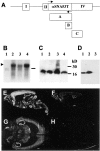
Fig. 1.
Fig. 1.
Transgene expression. A, Schematic representation of the transgene. Roman numeralsrefer to exons in the endogenous murine Thy1 gene. Boxesrepresent a complete α-synuclein coding cDNA probe (A), a C-terminal 111 bp α-synuclein cDNA probe (B), and a Thy1 3′ untranslated region probe (C) (Lüthi et al., 1997).Arrows represent PCR primers for genotyping.B, Northern blot analysis of 10 μg of total brain RNA using probe A and brain RNA of a C57BL/6 nontransgenic mouse (lane 1), a line 9813 mouse (lane 2), a line 9956 mouse (lane 3), and the single transgenic male of line 9832 (lane 4). Thearrowhead refers to the transgene mRNA, and thehorizontal bar marks the position of endogenous mouse α-synuclein mRNA. C, Immunoblot analysis showing increased levels of α-synuclein protein in brain homogenates of a representative mouse of each of the lines 9956 (lane 1), 9813 (lane 2), the single F1 male of line 9832 (lane 3), and C57BL/6 (lane 4). The polyclonal rabbit antibody used (Chemicon) detects both mouse and human α-synuclein protein. D, Immunoblot using the antibody LB509 (specifically detects human but not the mouse α-synuclein protein; Zymed) and line 9956 (lane 1), line 9813 (lane 2), and C57BL/6 (lane 3).E, F, In situhybridization in sagittal brain sections of a line 9813 mouse (E) and a C57BL/6 mouse (F) using 35S-labeled cRNA corresponding to probe A (A). G, H, In situ hybridization in horizontal brain sections of a line 9813 mouse (G) and a C57BL/6 mouse (H) using 35S-labeled cRNA corresponding to probe B (A).
Fig. 2.
Fig. 2.
Rotating rod performance of transgenic mice. Starting at age 40 d and up to age 200 d, transgenic (n = 7) and C57BL/6 nontransgenic (n = 12) littermate male mice were tested for endurance to stay on the rotating rod. The mice were tested weekly, and their performance is shown for two different rotation speeds, speed 1 (12 rpm) and speed 3 (36 rpm). C57BL/6 mice showed maximum endurance performance (60 sec) at both speeds and as shown for speed 3 (36 rpm).
Fig. 3.
Fig. 3.
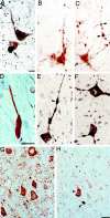
Lewy-like pathology in the transgenic mouse spinal cord. A–H correspond to sections through the anterior horn. A, Prominent perikaryal and proximal neuritic α-synuclein staining of motor neurons in a transgenic mouse spinal cord section but not in C57BL/6 (B).C, Campbell–Switzer-stained motor neurons in a transgenic mouse spinal cord, which also immunoreacted with anti-ubiquitin antibody (D). E,G, Anti-GFAP and anti-phosphotyrosine antibody stainings (F, H) in transgenic (E, F) versus nontransgenic (G, H) C57BL/6 spinal cord show evidence for astrocytic gliosis (E) and reactive microglia (F) specific to the transgenic tissue. Scale bars: A–D (in D), 20 μm;E–H (in H), 20 μm. Magnification: A–D, 250×; E–H, 200×.
Fig. 4.
Fig. 4.
Neuromuscular degeneration. A, Longitudinal section of a spinal root with human α-synuclein-immunoreactive (LB509 antibody) nerve fibers that are specific to the transgenic mice (C57BL/6 not shown). B, Holmes–Luxol-stained spinal root showing axonal degeneration with breakdown and segmentation of myelin into ellipsoids (“digestive chambers”). C, Cross-sectioned bundle of nerve fibers in a muscle showing strongly human α-synuclein-specific (antibody LB509) immunoreactive axons. D, Cross-sectioned muscle fiber bundle containing small angular fibers consistent with denervation (arrowheads), indicating neurogenic muscle atrophy (Holmes–Luxol stain). Scale bars:A–D, 20 μm. E–J, Medial gastrocnemius NMJs from C57BL/6 (E) and two different line 9813 mice (mouse 1, F–H; mouse 2, I,J), aged 6.5 months. The combined silver-esterase reaction reveals nerves in black and the synaptic acetylcholine esterase reaction product (asterisks) inblue. NMJs in the transgenic mice show neuropathological changes ranging from swellings of preterminal nerves (F, arrow), thinning out of the preterminal nerve (G), retraction of the nerve from the synaptic region (H), to complete denervation (I, arrows). A thin and characteristically twisted nerve sprout (arrows) has regrown to a denervated synapse shown in J. Dark ovals (in I and J) are attributable to background labeling of nuclei. Scale bar, 40 μm. Magnification: A, 100×; B,C, 400×; D, 200×; E–J, 400×.
Fig. 5.
Fig. 5.
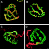
Neurodegeneration of soleus neuromuscular junctions. AD, The structure of the postsynaptic apparatus is visualized by α-bungarotoxin staining of AChRs (green), which was similar in C57BL/6 (A, C) and transgenic (B,D) mice. Costainings are shown for α-bungarotoxin (green) and synaptophysin (A,B), and α-bungarotoxin and neurofilament (C, D). Scale bar, 3 μm. Magnification, 700×.
Fig. 6.
Fig. 6.
α-Synuclein protein expression in the transgenic mouse brain. A–F, α-Synuclein staining in the hippocampal CA1 region (A, B) and neocortex (D, E) of a transgenic mouse compared with the respective regions in a nontransgenic C57BL/6 mouse (C, F). Unlike in the transgenic brain, the C57BL/6 hippocampus lacks α-synuclein immunoreactivity in CA1 pyramidal cell bodies in stratum pyramidale (st.p.). In both mice, immunostaining is prominent in stratum radiatum (st.r.). Strongly immunoreactive neurites (i.e., CA1 cell dendrites) in stratum radiatum are specific to the transgenic brain. Similar dendritic structures in the C57BL/6 brain appear pale and without α-synuclein. Immunostaining was in paraffin (A, D; human α-synuclein-specific antibody LB509) and free-floating brain sections (B,C, E, F; antibody detects both mouse and human α-synuclein). Scale bars: A,D (in D), 20 μm (250× magnification);B, C, E, F (in F), 20 μm (200× magnification).
Fig. 7.
Fig. 7.
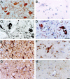
α-Synuclein and ubiquitin in transgenic mouse and human PD brain. A–F, Sections are shown of a human PD substantia nigra (A, D) and a transgenic mouse brain pontine reticular nucleus (B,C, E, F) immunostained for α-synuclein (A–C) and ubiquitin (D–F). Note the prominent somatodendritic staining for α-synuclein and the neuritic staining for ubiquitin in both human and mouse neurons. Lewy-like neurites are indicated by arrowheads. A cell of the substantia nigra in the human PD brain shows a Lewy body inclusion (A,arrow). G, H, Two consecutive 3 μm paraffin sections of the same group of cells in a cerebellar nucleus, stained for α-synuclein (LB509 antibody;G) and ubiquitin (H).G, Several neurons showing similar degrees of perikaryal α-synuclein immunoreactivity. Many cross-sectioned neurites are also stained. H, Abundant ubiquitin immunoreactivity is seen in only one of the three grouped cells and a nearby neurite (arrowheads in H and Gindicate the same cell and neurite showing α-synuclein immunoreactivity). In addition, compared with the number of α-synuclein-positive neurites in G, inH a smaller number of these structures (dark and punctate) show immunoreactivity for ubiquitin. Scale bars:A, D, 20 μm (magnification:A, 320×; D, 630×); B, C, E–H (in H), 20 μm (magnification, 400×).
Fig. 8.
Fig. 8.
Ultrastructural features of neurons in Thy1αSNA53T mice. Shown are immunoelectron (A,B) and a conventional micrograph (C) of spinal cord gray matter. A,B, Five-month-old Thy1αSNA53T mouse of line 9813. The immunoelectron micrographs are specifically stained for human but not mouse α-synuclein (LB509 antibody). A, Longitudinal section through a small dendritic appendage showing prominent staining of elements in the dendritic cytoplasm. B, Immunolabeled cross-sectioned dendrite, showing the fine granular composition of the α-synuclein-containing structures, which occasionally appeared attached to the endoplasmic reticulum and mitochondria, for example.C, Conventionally stained electron micrograph of a brainstem section of a 4-month-old transgenic mouse showing fine granular electron-dense material in dendritic profiles, most evident in the cross-sectioned dendrite located in the top rightpart between the three larger and a smaller myelinated axon. Scale bars: A, 1.7 μm (4500× magnification);B, 0.6 μm (12,000× magnification); C, 1.9 μm (5000× magnification).
Fig. 9.
Fig. 9.
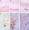
Immunopathology in Thy1αSNwt.A, Cerebral cortex showing prominent somatic and axodendritic α-synuclein immunostaining on neurons of the deep cortical layers. B, CA1 region of the hippocampus showing prominent α-synuclein immunostaining in principal cell somata and dendrites. C, CA3 sector of the hippocampus. Note some pyramidal cells with prominent cytoplasmic staining next to unstained cells and the immunostaining of the mossy fiber terminals.D, Deep mesencephalic nucleus with a neuron showing a strong α-synuclein immunostaining in soma and proximal dendrites. Note the presence of unstained axons in the vicinity.E, Spinal cord longitudinal section with α-synuclein-immunostained motor neurons and enlarged proximal dendrites (arrow). F, Same area as inE showing ubiquitin immunostaining in the cytoplasm of some neurons. Scale bars: A, B (inB), 20 μm; C, D (inD), 20 μm; E, F (inF), 20 μm. Magnification: A,B, 100×; C, D, 250×;E, F, 320×.

References

    1. Aigner L, Arber S, Kapfhammer JP, Laux T, Schneider C, Botteri F, Brenner HR, Caroni P. Overexpression of the neuronal growth-associated protein GAP-43 induces nerve sprouting in the adult nervous system of transgenic mice. Cell. 1995;83:269–278. - PubMed
    1. Arai T, Ueda K, Ikeda K, Akiyama H, Haga C, Kondo H, Kuroki N, Niizato K, Iritani S, Tsuchiya K. Argyrophilic glial inclusions in the midbrain of patients with Parkinson's disease and diffuse Lewy body disease are immunopositive for NACP/alpha-synuclein. Neurosci Lett. 1999;259:83–86. - PubMed
    1. Arima K, Uéda K, Sunohara N, Hirai S, Izumiyama Y, Tonozuka-Uehara H, Kawai M. Immunoelectron microscopic demonstration of NACP/α-synuclein-epitopes on the filamentous components of Lewy bodies in Parkinson's disease and in dementia with Lewy bodies. Brain Res. 1998;808:93–100. - PubMed
    1. Baba M, Nakajo S, Tu PH, Tomita T, Nakaya K, Lee VM, Trojanowski JQ, Iwatsubo T. Aggregation of α-synuclein in Lewy bodies of sporadic Parkinson's disease and Dementia with Lewy bodies. Am J Pathol. 1998;152:879–884. - PMC - PubMed
    1. Bennett MC, Bishop JF, Leng Y, Chock PB, Chase TN, Mouradian MM. Degradation of alpha-synuclein by proteasome. J Biol Chem. 1999;274:33855–33858. - PubMed

MeSH terms