Review
. 1999 Aug;1(3):208-19.
doi: 10.1038/sj.neo.7900034.

"...those left behind." Biology and oncology of invasive glioma cells

Affiliations
Review

"...those left behind." Biology and oncology of invasive glioma cells

M E Berens et al. Neoplasia. 1999 Aug.

Abstract

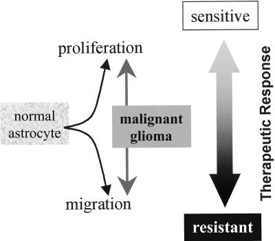
Although significant technical advances in surgical and radiation treatment for brain tumors have emerged in recent years, their impact on clinical outcome for patients has been disappointing. A fundamental source of the management challenge presented by glioma patients is the insidious propensity of the malignant cells to invade into adjacent normal brain. Invasive tumor cells escape surgical removal and geographically dodge lethal radiation exposure. Recent improved understanding of the biochemistry and molecular determinants of glioma cell invasion provide valuable insight to the underlying biological features of the disease, as well as illuminating possible new therapeutic targets. Heightened commitment to migrate and invade is accompanied by a glioma cell's reduced proliferative activity. The microenvironmental manipulations coincident to invasion and migration may also impact the glioma cell's response to cytotoxic treatments. These collateral aspects of the glioma cell invasive phenotype should be further explored and exploited as novel antiglioma therapies.


Figures

Figure 1
Figure 1
Mortality rates for primary malignant brain tumors in the United States for the 20-year interval 1973 to 1993. No significant appreciable improvement in these mortality rates has been made in these two decades, highlighting the difficulty in therapeutic advances for brain tumor patients. (Symbols: Diamond, white men; square, white women; triangle, black men; and circle, black women) (Adapted from Ref. (171); with permission).
Figure 2
Figure 2
Relationship between extent of GBM removed by surgery and patient survival. Survival curves illustrate outcome for 172 patients receiving extensive surgical resection (circles), 301 patients receiving partial resection (triangles), and 130 patients having biopsy procedures (squares) as the sole treatment for GBM. The greater the extent of tumor removed, the longer patients survive. GBM is a lethal disease irrespective of how aggressive the management strategy becomes. (Adapted from Ref. (172); with permission).
Figure 3
Figure 3
Histologic appearance of infiltrating edge from high grade astrocytoma. Two different specimens are evaluated at low power (A and C) and high power (B and D) microscopy. The first case (A and B) shows diffusive infiltration of tumor cells into the brain parenchyma (B), with evidence of a very gradual gradient of declining tumor cell density moving left-to-right in the field. The second specimen (C and D) presents with a cellular tumor core and a well-delineated tumor rim (C), but at higher magnification (D) the centripetal dissemination of tumor cells can be seen. Typical glioma cell morphological heterogeneity is also evident in the higher magnification images.
Figure 4
Figure 4
Schematic diagram of modifications in gene expression (loss or gain) that promote the invasive phenotype of glioma cells. The microenvironment of astrocytes, oligodendrocytes, and ECM is the backdrop against which opportunistic or tumor-derived reactions lead to glioma cell locomotion. The overview depicts the various reported determinants of invasion gleaned from reports on the different receptors or proteins and does not intend to infer that the changes are linked, sequential or concordant. It is likely that glioma cells use common mechanisms of invasion, which include detachment consequences as well as attachment events in their successful penetration into brain parenchyma. The biochemical determinants of invasion into white matter are not necessarily the same as those needed for invasion in the perivascular space.
Figure 5
Figure 5
Proliferation index of stationary (A) and migratory (B) human glioma cells assessed by immunocytochemistry for MIB-1. Cells were deposited as confluent discs of cells (74) onto substrates of tissue culture-treated glass or laminin that were blocked with bovine serum albumin, then the population of cells was monitored for lateral dissemination from the initial site. Cells were fixed after 48 hours of culture, probed with anti-MIB-1 antibodies (DAKO, Carpinteria, CA), treated with biotinylated anti-mouse secondary antibodies (Pierce, Rockford, IL), then incubated with strepavidin-HRP (Amersham Pharmacia Biotech, Piscataway, NJ). Reaction with DAB produces a brown insoluble product where the primary antibody bound MIB-1. Cells on glass (A) manifest minimal migration evidenced by the small intercellular space and the tightness of the perimeter cells to one another. The population of nonmotile cells is uniformly labeled by anti-MIB-1, indicating that the population of cells is largely in cell cycle. Cells on laminin (B) were in a sustained migration mode evidenced by the increased intercellular spaces and the dispersion of the perimeter cells away from the initial site of seeding. These motile cells, especially those at the periphery, show greatly diminished proliferation commitment.
Figure 6
Figure 6
Oncology of invasive glioma cells. A dichotomy exists between proliferation commitment and the migratory phenotype of normal and malignant glial cells. Arrested migration leads to more proliferation, whereas suppressed proliferation shifts glioma cells to a more migratory phenotype. Highly migratory glioma cells also show a relative resistance to cytotoxic insult (chemotherapy or radiation therapy), whereas nonmigratory glioma cells in the proliferative pool are more responsive to these treatments. Recognition of the role migration has on both proliferation and response to therapy, and identification of means by which to manipulate motility behavior to exploit therapeutic gain, should provide new opportunities for brain tumor treatments.

References

    1. 1997 Annual Report. Central Brain Tumor Registry of the United States Chicago, IL. 1998:1.
    1. Burger PC, Dubois PJ, Schold SC, Smith KR, Odom GL, Crafts DC, Giangaspero F. Computerized tomographic and pathologic studies of the untreated, quiescent, and recurrent glioblastoma multiforme. J Neurosurg. 1983;58:159–169. - PubMed
    1. Gaspar LE, Fisher BJ, MacDonald DR, LeBer DV, Halperin EC, Schold SC, Cairncross JG. Supratentorial malignant glioma: patterns of recurrence and implications for external beam local treatment. Int J Radiat Oncol Biol Phys. 1992;24:55–57. - PubMed
    1. Brem H, Ewend MG, Piantadosi S, Greenhoot J, Burger PC, Sisti M. The safety of interstitial chemotherapy with BCNU-loaded polymer followed by radiation therapy in the treatment of newly diagnosed malignant gliomas: Phase I trial. J Neurooncol. 1995;26:111–123. - PubMed
    1. Ram Z, Culver KW, Oshiro EM, Viola JJ, DeVroom HL, Otto E, Long Z, Chiang Y, McGarrity GJ, Muul LM, Katz D, Blaese RM, Oldfield EH. Therapy of malignant brain tumors by intratumoral implantation of retroviral vector-producing cells [see comments] Nat Med. 1997;3:1354–1361. - PubMed

Publication types

Substances

LinkOut - more resources