Differentiation of human malignant melanoma cells that escape apoptosis after treatment with 9-nitrocamptothecin in vitro
- PMID: 10935478
- PMCID: PMC1508080
- DOI: 10.1038/sj.neo.7900025
Differentiation of human malignant melanoma cells that escape apoptosis after treatment with 9-nitrocamptothecin in vitro
Abstract
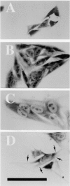
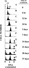
After in-vitro exposure to 0.05 micromol/L 9-nitrocamptothecin (9NC) for periods of time longer than 5 days, 65% to 80% of the human malignant melanoma SB1B cells die by apoptosis, whereas the remaining cells are arrested at the G2-phase of the cell cycle. Upon discontinuation of exposure to 9NC the G2-arrested cells resume cell cycling or remain arrested depending on the duration of 9NC exposure. In contrast to cycling malignant cells, the cells irreversibly arrested at G2 exhibit features of normal-like cells, the melanocytes, as assessed by the appearance of dendrite-like structures; loss of proliferative activity; synthesis of the characteristic pigment, melanin; and, particularly, loss of tumorigenic ability after xenografting in immunodeficient mice. Further, the expression of the cyclin-dependent kinase inhibitor p16 is upregulated in the 9NC-treated, G2-arrested, but downregulated in density G1-arrested cells, whereas the reverse is observed in the expression of another cyclin-dependent kinase inhibitor, p21. These results suggest that malignant melanoma SB1B cells that escape 9NC-induced death by apoptosis undergo differentiation toward nonmalignant, normal-like cells.
Figures


Similar articles
-
Induction of senescent cell-derived inhibitor of DNA synthesis gene, SDI1, in hepatoblastoma (HepG2) cells arrested in the G2-phase of the cell cycle by 9-nitrocamptothecin.Lab Invest. 1995 Jul;73(1):118-27. Lab Invest. 1995. PMID: 7603034
-
Cytotoxic efficacy of 9-nitrocamptothecin in the treatment of human malignant melanoma cells in vitro.Cancer Res. 1994 Feb 1;54(3):771-6. Cancer Res. 1994. PMID: 8306340
-
Regression of human breast carcinoma tumors in immunodeficient mice treated with 9-nitrocamptothecin: differential response of nontumorigenic and tumorigenic human breast cells in vitro.Cancer Res. 1993 Apr 1;53(7):1577-82. Cancer Res. 1993. PMID: 8453626
-
Camptothecin and 9-nitrocamptothecin (9NC) as anti-cancer, anti-HIV and cell-differentiation agents. Development of resistance, enhancement of 9NC-induced activities and combination treatments in cell and animal models.Anticancer Res. 2003 Sep-Oct;23(5A):3623-38. Anticancer Res. 2003. PMID: 14666658 Review.
-
Cytometry of cyclin proteins.Cytometry. 1996 Sep 1;25(1):1-13. doi: 10.1002/(SICI)1097-0320(19960901)25:1<1::AID-CYTO1>3.0.CO;2-N. Cytometry. 1996. PMID: 8875049 Review.
Cited by
-
PPAR gamma regulates MITF and beta-catenin expression and promotes a differentiated phenotype in mouse melanoma S91.Pigment Cell Melanoma Res. 2008 Jun;21(3):388-96. doi: 10.1111/j.1755-148X.2008.00460.x. Epub 2008 Apr 26. Pigment Cell Melanoma Res. 2008. PMID: 18444964 Free PMC article.
-
Targeting p53 for Melanoma Treatment: Counteracting Tumour Proliferation, Dissemination and Therapeutic Resistance.Cancers (Basel). 2021 Apr 1;13(7):1648. doi: 10.3390/cancers13071648. Cancers (Basel). 2021. PMID: 33916029 Free PMC article.
References
-
- Nathan FE, Berd D, Mastrangelo MJ. Chemotherapy of melanoma. In: Perry MC, editor. The Chemotherapy Source Book. 2nd edn. Baltimore, MD: Williams & Wilkins; 1996. pp. 1043–1069.
-
- Houghton A. Treatment for advanced melanoma. In: Balch CM, Houghton AN, Milton GW, Sober AJ, Soong S-Y, editors. Cutaneous Melanoma. Philadelphia, PA: Lippincott; 1992. pp. 468–497.
-
- Houghton AN, Legha S, Bajorin DF. Chemotherapy for metastatic melanoma. In: Balch CM, Houghton AN, Milton GW, Sober AJ, Soong S-Y, editors. Cutaneous Melanoma. Philadelphia, PA: Lippincott; 1992. pp. 498–508.
-
- Herlyn M, Houghton AN. Biology of melanocytes and melanoma. In: Balch CM, Houghton AN, Milton GW, Sober AJ, Soong S-Y, editors. Cutaneous Melanoma. Philadelphia, PA: Lippincott; 1992. pp. 82–92.
-
- Valyi-Nagy I, Shih IM, Gyorfi T, Greenstein D, Juhasz I, Elder DE, Herlyn M. Spontaneous and induced differentiation of human melanoma cells. Int J Cancer. 1993;54:159–165. - PubMed
Publication types
MeSH terms
Substances
LinkOut - more resources
Full Text Sources
Medical
