. 2000 Sep;20(17):6390-8.
doi: 10.1128/MCB.20.17.6390-6398.2000.

Functional studies on the candidate ATPase domains of Saccharomyces cerevisiae MutLalpha

Affiliations

Functional studies on the candidate ATPase domains of Saccharomyces cerevisiae MutLalpha

P T Tran et al. Mol Cell Biol. 2000 Sep.

Abstract

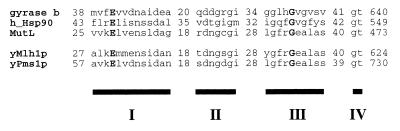
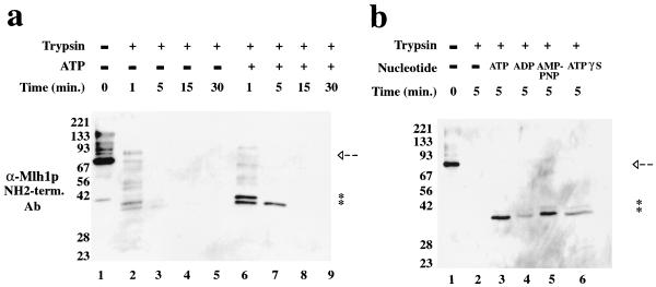
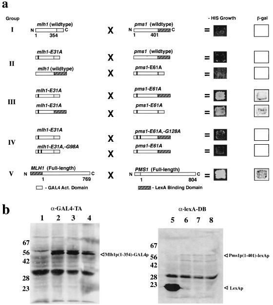
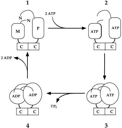
Saccharomyces cerevisiae MutL homologues Mlh1p and Pms1p form a heterodimer, termed MutLalpha, that is required for DNA mismatch repair after mismatch binding by MutS homologues. Recent sequence and structural studies have placed the NH(2) termini of MutL homologues in a new family of ATPases. To address the functional significance of this putative ATPase activity in MutLalpha, we mutated conserved motifs for ATP hydrolysis and ATP binding in both Mlh1p and Pms1p and found that these changes disrupted DNA mismatch repair in vivo. Limited proteolysis with purified recombinant MutLalpha demonstrated that the NH(2) terminus of MutLalpha undergoes conformational changes in the presence of ATP and nonhydrolyzable ATP analogs. Furthermore, two-hybrid analysis suggested that these ATP-binding-induced conformational changes promote an interaction between the NH(2) termini of Mlh1p and Pms1p. Surprisingly, analysis of specific mutants suggested differential requirements for the ATPase motifs of Mlh1p and Pms1p during DNA mismatch repair. Taken together, these results suggest that MutLalpha undergoes ATP-dependent conformational changes that may serve to coordinate downstream events during yeast DNA mismatch repair.


Figures

FIG. 1
FIG. 1
NH2-terminal ATPase domains of GHL ATPases. ATPase motifs I to IV are designated by black boxes, and sequences are shown above motif boxes. Numbers correspond to the number of amino acids preceding or following sequence alignments. Boldface letters are the absolutely conserved residues that were substituted for with alanine in Mlh1p and Pms1p.
FIG. 2
FIG. 2
Adenine nucleotides alter trypsin sensitivity of MutLα. (a) One hundred fifty nanograms of MutLα was subjected to proteolysis with modified trypsin as described in Materials and Methods in the presence or absence of 5 mM ATP for the indicated time at 30°C. Products were treated with SDS-sample buffer, boiled, separated on an SDS-PAGE (10% polyacrylamide) gel, and detected by immunoblotting with anti-4×His antibody. Arrows denote full-length 6×His-Mlh1p, and asterisks designate NH2-terminal (term.) fragments of 6×His-Mlh1p that are protected from proteolysis in the presence of ATP. Equal loading of samples and even transfer of the blot were demonstrated by using a polyclonal antibody raised against the COOH terminus of Mlh1p (data not shown). (b) The same analysis was performed as described for panel a, but the effects of 5 mM adenine nucleotides ADP, AMP-PNP, and ATPγS were examined.
FIG. 3
FIG. 3
Two-hybrid analysis detects NH2-terminal Mlh1p and Pms1p interaction. (a) Boxes correspond to bait and prey constructs tested for interaction. The residues included in the fusions are indicated below the group I and V constructs, respectively. Amino acid substitutions designated above each construct are indicated by black bars within the construct boxes. Interaction is scored as growth on −HIS media and blue color development with the substrate X-Gal (5-bromo-4-chloro-3-indolyl-β-d-galactopyranoside) as described in Materials and Methods. Group I, wild-type NH2-terminal fusion fragments; group II, one NH2-terminal fusion fragment contains a hydrolysis point mutation; group III, both NH2-terminal fusion fragments contain hydrolysis point mutations; group IV, one NH2-terminal fusion fragment has the indicated compound mutations; and group V, positive control reaction with full-length Pms1p and Mlh1p. (b) Western analysis of L40 strains with two-hybrid constructs from panel a using anti-GAL4-TA or anti-lexA-DB monoclonal antibody as described in Materials and Methods. Lanes: 1, pCAD3 (empty vector); 2, pCAD-mlh1 N-354; 3, pCAD-mlh1-E31A N-354; 4, pCAD-mlh1-E31A, -G98A N-354; 5, pNBTM (lexA); 6, pNBTM-pms1 N-401; 7, pNBTM-pms1-E61A N-401; 8, pNBTM-pms1-E61A, -G128A N-401. Fusion products and lexAp are indicated by arrowheads. The approximately 90-kDa band in lanes 1 to 4 may be endogenous Gal4p. The other bands present in control lanes 1 and 5 and in lanes 2 to 4 and 6 to 8, respectively, represent nonspecificity by the primary and secondary antibodies. In lanes 6 to 8, the faster-migrating specific anti-lexA-DB reacting species is unknown, but may be a pms1p(1-401)-lexAp degradation product.
FIG. 4
FIG. 4
A model for the yeast MutLα ATPase cycle. Briefly, intermediate 1 is the nucleotide-free state. ATP binding induces conformational changes in the NH2 termini of Mlh1p and Pms1p, represented by a change in shape from rectangular to oval that occurs in the step(s) between intermediates 2 and 3. Intermediate 3 is heterodimerization of the NH2 termini of Mlh1p and Pms1p in the ATP-bound state. Intermediate 4 is the ADP-bound form following ATP hydrolysis. The mlh1-G98A and pms1-G128A ATP-binding mutants were constructed to affect the transition(s) from intermediate 1 to intermediate 2 and/or intermediate 2 to intermediate 3. In contrast, the ATP hydrolysis mutations, mlh1-E31A and pms1-E61A, were modeled to prevent the transition from intermediate 3 to intermediate 4. M, the NH2 terminus of Mlh1p; P, the NH2 terminus of Pms1p; C, COOH termini of Mlh1p and Pms1p. Each arrow may represent multiple distinct steps. This model, which is consistent with the studies reported here, was adapted from a model for MutL proposed by Ban and Yang (9).

References

    1. Alani E, Cao L, Kleckner N. A method for gene disruption that allows repeated use of URA3 selection in the construction of multiply disrupted yeast strains. Genetics. 1987;116:541–545. - PMC - PubMed
    1. Alani E, Chi N-W, Kolodner R. The Saccharomyces cerevisiae Msh2 protein specifically binds to duplex oligonucleotides containing mismatched DNA base pairs and insertions. Genes Dev. 1995;9:234–247. - PubMed
    1. Alani E, Sokolsky T, Studamire B, Miret J J, Lahue R S. Genetic and biochemical analysis of Msh2p-Msh6p: role of ATP hydrolysis and Msh2p-Msh6p subunit interactions in mismatch base pair recognition. Mol Cell Biol. 1997;17:2436–2447. - PMC - PubMed
    1. Ali J A, Jackson A P, Howells A J, Maxwell A. The 43-kilodalton N-terminal fragment of the DNA gyrase b protein hydrolyzes ATP and binds coumarin drugs. Biochemistry. 1993;32:2717–2724. - PubMed
    1. Aronshtam A, Marinus M G. Dominant negative mutator mutations in the mutL gene of Escherichia coli. Nucleic Acids Res. 1996;24:2498–2504. - PMC - PubMed

Publication types

MeSH terms