. 2000 Sep;20(17):6476-82.
doi: 10.1128/MCB.20.17.6476-6482.2000.

The Rad51 paralog Rad51B promotes homologous recombinational repair

Affiliations

The Rad51 paralog Rad51B promotes homologous recombinational repair

M Takata et al. Mol Cell Biol. 2000 Sep.

Abstract

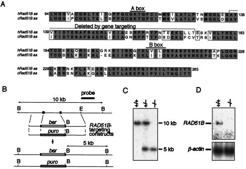
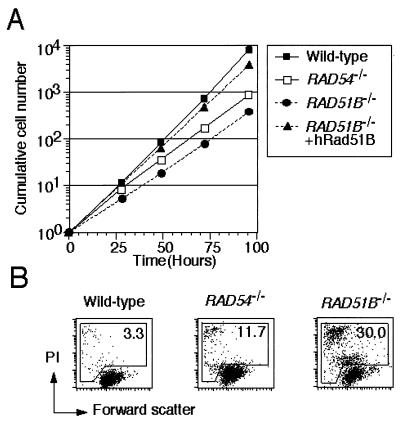
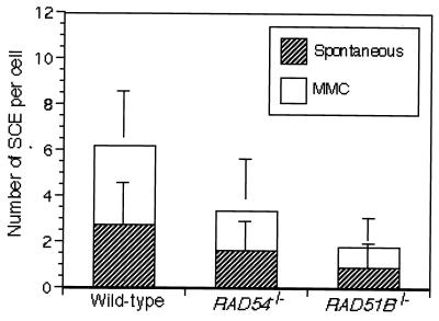
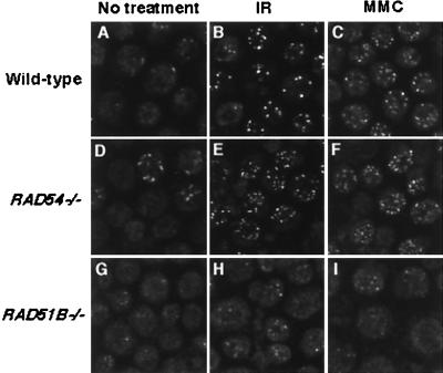
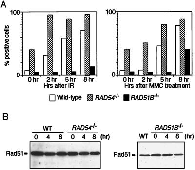
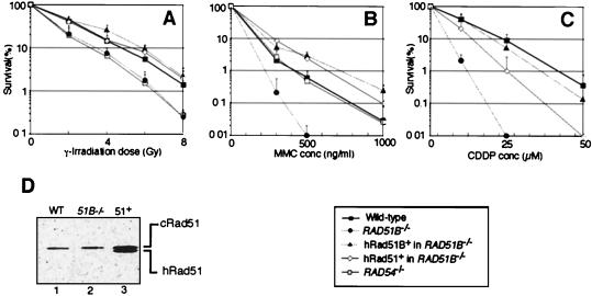
The highly conserved Saccharomyces cerevisiae Rad51 protein plays a central role in both mitotic and meiotic homologous DNA recombination. Seven members of the Rad51 family have been identified in vertebrate cells, including Rad51, Dmc1, and five Rad51-related proteins referred to as Rad51 paralogs, which share 20 to 30% sequence identity with Rad51. In chicken B lymphocyte DT40 cells, we generated a mutant with RAD51B/RAD51L1, a member of the Rad51 family, knocked out. RAD51B(-/-) cells are viable, although spontaneous chromosomal aberrations kill about 20% of the cells in each cell cycle. Rad51B deficiency impairs homologous recombinational repair (HRR), as measured by targeted integration, sister chromatid exchange, and intragenic recombination at the immunoglobulin locus. RAD51B(-/-) cells are quite sensitive to the cross-linking agents cisplatin and mitomycin C and mildly sensitive to gamma-rays. The formation of damage-induced Rad51 nuclear foci is much reduced in RAD51B(-/-) cells, suggesting that Rad51B promotes the assembly of Rad51 nucleoprotein filaments during HRR. These findings show that Rad51B is important for repairing various types of DNA lesions and maintaining chromosome integrity.


Figures

FIG. 1
FIG. 1
Gene targeting of RAD51B loci. (A) Amino acid (aa) sequence comparison between human and chicken Rad51B cDNAs. The Walker A and B motifs for nucleotide binding are overlined, and the sequence deleted by gene targeting is indicated. Letters in gray boxes represent identical amino acids in the two species, whereas those in open boxes represent similar (P, A, G, S, and T; E, D, N, and Q; V, I, L, and M; F, W, and Y; R, K, and H) amino acids. Numbers denote amino acid positions. (B) Schematic representation of part of the RAD51B locus, the gene disruption constructs, and the configuration of the targeted alleles. B, BamHI site; E, EcoRI site. Solid boxes indicate the positions of the exons. Only disrupted exons are indicated. (C) Southern blot analysis of BamHI-digested genomic DNA from cells with the indicated genotypes of the RAD51B gene with the probe shown in panel B. The positions and sizes of the hybridizing fragments of the wild-type and targeted loci are indicated. (D) Northern blot analysis of total RNA with a chicken RAD51B cDNA fragment as a probe. The same membrane was reprobed with the chicken β-actin fragment (12).
FIG. 2
FIG. 2
Growth rate and viability of RAD51B−/− cells. (A) Growth curves of cells of the indicated genotypes. Means ± standard deviations of triplicate cultures are shown. (B) The level of spontaneous cell death was assessed by flow cytometric analysis of propidium iodide uptake and forward scatter representing cell size. The values shown are percentages of dead (propidium iodide-bright and propidium iodide-dim) cells.
FIG. 3
FIG. 3
Level of SCE per cell. Cells were incubated with or without MMC (0.05 μg/ml) for 12 h and treated with colcemid during the last 1.5 h of this incubation to enrich mitotic cells. One hundred fifty cells were analyzed in each preparation. Error bars represent standard errors calculated as previously described (58).
FIG. 4
FIG. 4
Immunofluorescent visualization of Rad51 subnuclear foci. Wild-type (A to C), RAD54−/− (D to F), and RAD51B−/− (G to I) cells were analyzed 5 h after genotoxic treatments. Cells were treated with either 8 Gy of γ radiation (IR) (B, E, and H) or 500 ng of MMC per ml (for 1 h) (C, F, and I).
FIG. 5
FIG. 5
Induction of Rad51 focus-positive cells after genotoxic treatments. (A) Cells with the indicated genotypes were analyzed at the indicated time points after either γ irradiation (IR; 8 Gy) or MMC treatment (500 ng/ml, 1 h). A cell containing more than four distinct foci was scored as positive. Each bar represents the result of scoring at least 100 cells. (B) Rad51 protein expression before and after exposure to 8 Gy of γ irradiation. At the indicated time points, total cell lysates were prepared and the same amount of protein was loaded into each lane. WT, wild type.
FIG. 6
FIG. 6
Sensitivity of clones of the indicated genotypes to DNA-damaging agents. The fractions of the surviving colonies after the indicated treatment of cells compared to nontreated controls of the same genotype are shown on the y axis on a logarithmic scale. Panels: A, γ irradiation; B, MMC; C, cisplatin (CDDP). The dose of radiation treatment and concentrations (conc) of MMC and cisplatin are displayed on the x axis on a linear scale in each graph. The data shown are means ± standard deviations of at least three separate experiments. The cisplatin sensitivity data of RAD54−/− cells are not shown here but were previously described (65). (D) Western blot analysis with rabbit anti-Rad51 serum showing the amounts of endogenous chicken Rad51 and human Rad51 proteins derived from the transgene. Lanes: 1, wild-type (WT) DT40; 2, RAD51B−/− cells; 3, hRad51-expressing RAD51B−/− cells.

References

    1. Aboussekhra A, Chanet R, Adjiri A, Fabre F. Semidominant suppressors of Srs2 helicase mutations of Saccharomyces cerevisiae map in the RAD51 gene, whose sequence predicts a protein with similarities to procaryotic RecA proteins. Mol Cell Biol. 1992;12:3224–3234. - PMC - PubMed
    1. Albala J S, Thelen M P, Prange C, Fan W, Christensen M, Thompson L H, Lennon G G. Identification of a novel human RAD51 homolog, RAD51B. Genomics. 1997;46:476–479. - PubMed
    1. Basile G, Aker M, Mortimer R K. Nucleotide sequence and transcriptional regulation of the yeast recombinational repair gene RAD51. Mol Cell Biol. 1992;12:3235–3246. - PMC - PubMed
    1. Baumann P, West S C. Role of the human RAD51 protein in homologous recombination and double-stranded-break repair. Trends Biochem Sci. 1998;23:247–251. - PubMed
    1. Bezzubova O Y, Silbergleit A, Yamaguchi-Iwai Y, Takeda S, Buerstedde J M. Reduced X-ray resistance and homologous recombination frequencies in a RAD54−/− mutant of the chicken DT40 cell line. Cell. 1997;89:185–193. - PubMed

Publication types

MeSH terms