. 2000 Sep;182(17):4719-29.
doi: 10.1128/JB.182.17.4719-4729.2000.

RNase III processing of intervening sequences found in helix 9 of 23S rRNA in the alpha subclass of Proteobacteria

Affiliations

RNase III processing of intervening sequences found in helix 9 of 23S rRNA in the alpha subclass of Proteobacteria

E Evguenieva-Hackenberg et al. J Bacteriol. 2000 Sep.

Abstract

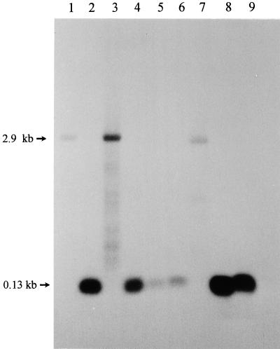
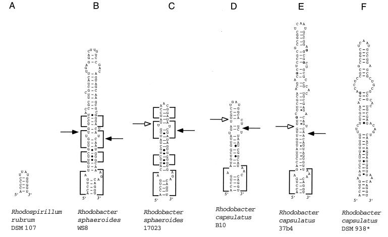
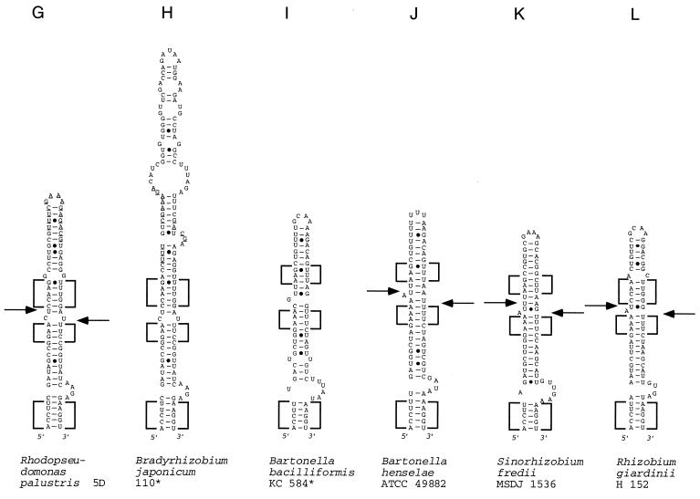
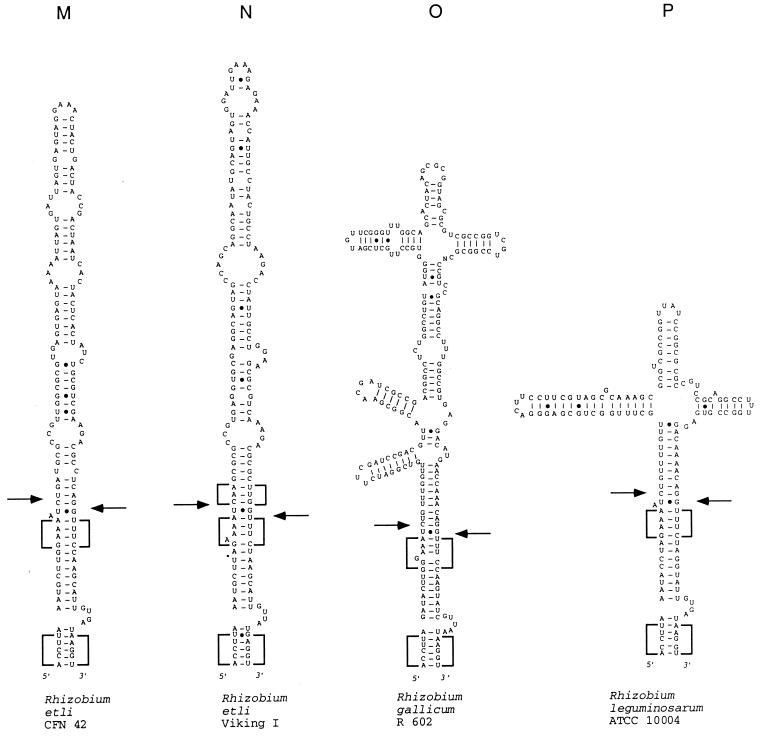
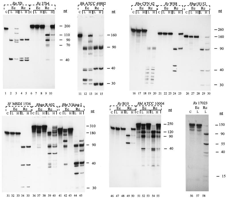
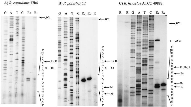
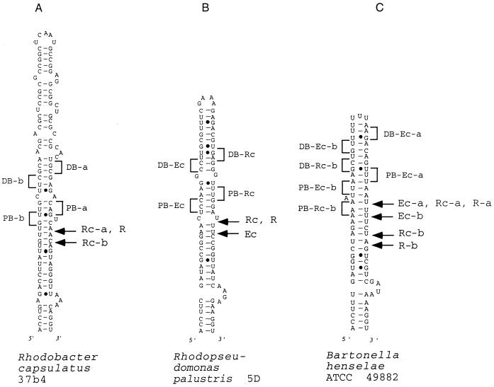
We provide experimental evidence for RNase III-dependent processing in helix 9 of the 23S rRNA as a general feature of many species in the alpha subclass of Proteobacteria (alpha-Proteobacteria). We investigated 12 Rhodobacter, Rhizobium, Sinorhizobium, Rhodopseudomonas, and Bartonella strains. The processed region is characterized by the presence of intervening sequences (IVSs). The 23S rDNA sequences between positions 109 and 205 (Escherichia coli numbering) were determined, and potential secondary structures are proposed. Comparison of the IVSs indicates very different evolutionary rates in some phylogenetic branches, lateral genetic transfer, and evolution by insertion and/or deletion. We show that the IVS processing in Rhodobacter capsulatus in vivo is RNase III-dependent and that RNase III cleaves additional sites in vitro. While all IVS-containing transcripts tested are processed in vitro by RNase III from R. capsulatus, E. coli RNase III recognizes only some of them as substrates and in these substrates frequently cleaves at different scissile bonds. These results demonstrate the different substrate specificities of the two enzymes. Although RNase III plays an important role in the rRNA, mRNA, and bacteriophage RNA maturation, its substrate specificity is still not well understood. Comparison of the IVSs of helix 9 does not hint at sequence motives involved in recognition but reveals that the "antideterminant" model, which represents the most recent attempt to explain the E. coli RNase III specificity in vitro, cannot be applied to substrates derived from alpha-Proteobacteria.


Figures

FIG. 1
FIG. 1
Presence of short rRNA corresponding to approximately the first 130 nt of the 23S rRNA in some alpha-Proteobacteria as shown by Northern hybridization of total RNA separated on a 1.2% agarose formaldehyde gel with the radioactively labeled oligonucleotide 23Sup130. Lanes: 1, E. coli JM109; 2, R. capsulatus 37b4; 3, R. capsulatus Fm65; 4, R. capsulatus Fm65 (pRK2fm1); 5, R. sphaeroides 17023; 6, R. palustris 5D; 7, R. rubrum DSM 107; 8, R. gallicum R602; 9, R. giardinii H152.
FIG. 2
FIG. 2
Models of potential secondary structure of helix 9 in the 23S rRNA primary transcript of the following strains (asterisks indicate sequences obtained from the data bank, the other sequences were determined in our laboratory, and EMBL accession numbers are in parentheses): A, R. rubrum DSM 107 (AJ251267); B, R. sphaeroides WS8 (AJ251261); C, R. sphaeroides 17023 (AJ251260); D, R. capsulatus B10 (AJ251256); E, R. capsulatus 37b4 (AJ251255); F, R. capsulatus DSM 938* (reference 11); G, R. palustris 5D (AJ251262); H, B. japonicum 110* (reference 17); I, B. bacilliformis KC 584* (reference 22); J, B. henselae ATCC 49882 (AJ251257); K, S. fredii MSDJ 1536 (AJ251258); L, R. giardinii H152 (AJ251263); M, R. etli CFN 42 (AJ251265); R. etli Viking I (AJ251266); O, R. gallicum R602 (AJ251259), R. leguminosarum ATCC 10004 (AJ251264). (B to F) Rhodobacter group of helices. Boxes with highly conservative base pair occupation, specific for this group, are indicated. The differences between the sequences shown in panels E and F are in boldface letters. (G to P) Rhizobium-Bradyrhizobium group of helices. Boxes with highly conservative base pair occupation, specific for this group, are indicated. In panels G and H, sequences of high similarity around the putative deletion and/or insertion site are underlined. Arrows indicate the approximate positions of the RNase III processing sites as determined by RNA fragment length estimation (Table 4). Arrows on the left side of the helices indicate 5′-processing sites; arrows on the right side of the helices indicate 3′-processing sites. Filled arrowheads indicate primary processing sites; empty arrowheads indicate secondary processing sites.
FIG. 2
FIG. 2
Models of potential secondary structure of helix 9 in the 23S rRNA primary transcript of the following strains (asterisks indicate sequences obtained from the data bank, the other sequences were determined in our laboratory, and EMBL accession numbers are in parentheses): A, R. rubrum DSM 107 (AJ251267); B, R. sphaeroides WS8 (AJ251261); C, R. sphaeroides 17023 (AJ251260); D, R. capsulatus B10 (AJ251256); E, R. capsulatus 37b4 (AJ251255); F, R. capsulatus DSM 938* (reference 11); G, R. palustris 5D (AJ251262); H, B. japonicum 110* (reference 17); I, B. bacilliformis KC 584* (reference 22); J, B. henselae ATCC 49882 (AJ251257); K, S. fredii MSDJ 1536 (AJ251258); L, R. giardinii H152 (AJ251263); M, R. etli CFN 42 (AJ251265); R. etli Viking I (AJ251266); O, R. gallicum R602 (AJ251259), R. leguminosarum ATCC 10004 (AJ251264). (B to F) Rhodobacter group of helices. Boxes with highly conservative base pair occupation, specific for this group, are indicated. The differences between the sequences shown in panels E and F are in boldface letters. (G to P) Rhizobium-Bradyrhizobium group of helices. Boxes with highly conservative base pair occupation, specific for this group, are indicated. In panels G and H, sequences of high similarity around the putative deletion and/or insertion site are underlined. Arrows indicate the approximate positions of the RNase III processing sites as determined by RNA fragment length estimation (Table 4). Arrows on the left side of the helices indicate 5′-processing sites; arrows on the right side of the helices indicate 3′-processing sites. Filled arrowheads indicate primary processing sites; empty arrowheads indicate secondary processing sites.
FIG. 3
FIG. 3
In vitro processing of transcripts containing helix 9 of 23S rRNA by R. capsulatus (Rc) and E. coli (Ec) RNase III at low and high monovalent ion concentrations. L, 130 mM KCl; H, 250 mM KCl; C, uncleaved substrate. Rp, R. palustris; Rc, R. capsulatus; Bh, B. henselae; Rhe, R. etli; Rs, R. sphaeroides; Rhgi, R. giardinii; Sf, S. fredi; Rhga, R. gallicum; Rhl, R. leguminosarum.
FIG. 4
FIG. 4
Primer extension analysis determines the rRNA 5′ ends obtained during in vitro cleavage of the transcripts from R. capsulatus 37b4 (A), R. palustris 5D (B), and B. henselae ATCC 49882 (C) with R. capsulatus (lanes Rc) and E. coli (lanes Ec) RNases III at the 3′-processing site in helix 9. The corresponding 5′ ends in vivo were detected by primer extension analysis using total RNA (lanes R) isolated from these strains. Lanes G, A, T, and C each refer to the corresponding nucleotide of the DNA template (cloned 23S rDNA region), as determined by sequencing. Parts of the in vitro transcripts and of the pre-rRNA sequences are indicated on the right side of each panel. The detected 5′ ends are marked by labeled arrows as follows: ⊘1, 5′ end of the 23S rRNA unprocessed in helix 9; ⊘2, 5′ end of the unprocessed in vitro transcript; Ec, 5′ end after RNase IIIEc cleavage in vitro; Rc, 5′ end after RNase IIIRc cleavage in vitro; R, 5′ end corresponding to the RNase III processing site in helix 9, detected in vivo; M, 5′ end arising from further maturation of rRNA in vivo.
FIG. 5
FIG. 5
Schematic representation of the RNase III 3′-processing sites in vitro and the in vivo 5′ ends found in helix 9 of 23S rRNA with primer extension analysis shown in Fig. 4. (A) R. capsulatus 37b. (B) R. palustris 5D. (C) B. henselae ATCC 49882. Labeled arrows: Ec, 5′ end after RNase IIIEc cleavage in vitro; Rc, 5′ end after RNase IIIRc cleavage in vitro; R, 5′ end corresponding to the RNase III processing site in helix 9, detected in vivo; PB and DB, proximal and distal boxes, respectively, found in the vicinity of the corresponding scissile bonds (compare with references and 38).

Similar articles

Cited by

References

    1. Amarger N, Macheret V, Laguerre G. Rhizobium gallicum sp. nov. and Rhizobium giardinii sp. nov., from Phaseolus vulgaris nodules. Int J Syst Bacteriol. 1997;47:996–1006. - PubMed
    1. Ausubel F M, Brent R, Kingston R E, Moore D D, Seidman J G, Smith J A, Struhl K. Current protocols in molecular biology. I. New York, N.Y: Greene Publishing Associates and Wiley-Interscience; 1989.
    1. Beringer J E. R-factor transfer in Rhizobium leguminosarum. J Gen Microbiol. 1974;84:188–198. - PubMed
    1. Burgin A B, Parodos K, Lane D J, Pace N R. The excision of intervening sequences from Salmonella 23S ribosomal RNA. Cell. 1990;60:405–414. - PubMed
    1. Conrad C, Rauhut R, Klug G. Different cleavage specificities of RNases III from Rhodobacter capsulatus and Escherichia coli. Nucleic Acids Res. 1998;26:4446–4453. - PMC - PubMed

Publication types

LinkOut - more resources