. 2000 Sep;182(17):4868-74.
doi: 10.1128/JB.182.17.4868-4874.2000.

Spy1, a histidine-containing phosphotransfer signaling protein, regulates the fission yeast cell cycle through the Mcs4 response regulator

Affiliations

Spy1, a histidine-containing phosphotransfer signaling protein, regulates the fission yeast cell cycle through the Mcs4 response regulator

K Aoyama et al. J Bacteriol. 2000 Sep.

Abstract

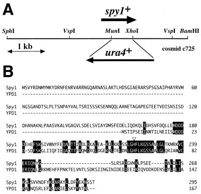
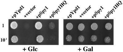
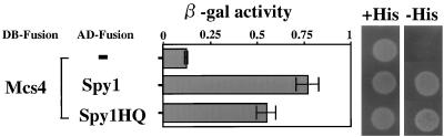
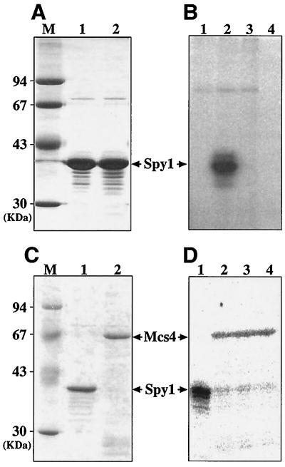
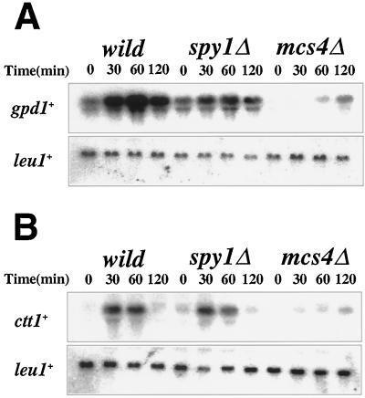
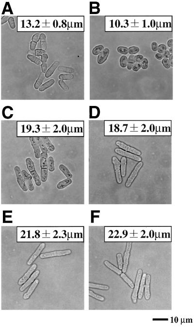
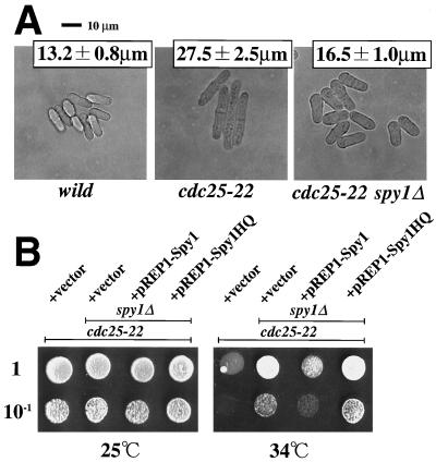
Common histidine-to-aspartate (His-to-Asp) phosphorelay signaling systems involve three types of signaling components: a sensor His kinase, a response regulator, and a histidine-containing phosphotransfer (HPt) protein. In the fission yeast Schizosaccharomyces pombe, two response regulators, Mcs4 and Prr1, have been identified recently, and it was shown that they are involved in the signal transduction implicated in stress responses. Furthermore, Mcs4 appears to be involved in mitotic cell-cycle control. However, neither the HPt phosphotransmitter nor His kinase has been characterized in S. pombe. In this study, we identified a gene encoding an HPt phosphotransmitter, named Spy1 (S. pombe YPD1-like protein). The spy1(+) gene showed an ability to complement a mutational lesion of the Saccharomyces cerevisiae YPD1 gene, which is involved in an osmosensing signal transduction. The result from yeast two-hybrid analysis indicated that Spy1 interacts with Mcs4. To gain insight into the function of Spy1, a series of genetic analyses were conducted. The results provided evidence that Spy1, together with Mcs4, plays a role in regulation of the G(2)/M cell cycle progression. Spy1-deficient cells appear to be precocious in the entry to M phase. In the proposed model, Spy1 modulates Mcs4 in a negative manner, presumably through a direct His-to-Asp phosphorelay, operating upstream of the Sty1 mitogen-activated protein kinase cascade.


Figures

FIG. 1
FIG. 1
The spy1+ gene encodes an HPt phosphotransmitter. (A) Schematic representation of the S. pombe chromosomal region encompassing the spy1+ gene. This region is carried in the c725 cosmid, whose entire nucleotide sequence has been determined (GenBank accession no. AL034352). In this study, an spy1Δ strain was constructed by inserting the ura4+ marker, as shown schematically. (B) The deduced amino acid sequence of Spy1 was aligned with that of the budding yeast Ypd1 protein. The open triangle indicates the presumed phosphorylation site (histidine). The amino acids identical in Spy1 and Ypd1 are highlighted.
FIG. 2
FIG. 2
The spy1+ gene is able to complement the mutational lesion of the YPD1 gene of S. cerevisiae. The ypd1Δ mutant carrying a plasmid harboring a composite PTP2 gene (PGAL1-PTP2) cannot grow on SC medium supplemented by glucose, while it can grow on SC medium supplemented with galactose (see the text) (+vector). Plasmid pKA014 carrying the recombinant spy1+ gene (designated as pSpy1) was transferred into the ypd1Δ mutant, and then the viability of the transformed cells (about 2 × 10² and 2 × 10³) was examined by spotting on galactose-synthetic complete (SC) medium (Gal) or glucose-SC medium (Glc). The cells were incubated for 3 days at 30°C. The same analyses were carried out for the plasmids carrying the mutant spy1 gene (designated as pSpy1HQ) as well as the budding yeast YPD1 gene (designated as pYpd1).
FIG. 3
FIG. 3
Yeast two-hybrid analysis. Two-hybrid analysis was carried out with the combination indicated. The results are shown as β-galactosidase activity and histidine auxotrophy (+His or −His). In this experiment, it should be noted that neither AD-Spy1 nor AD-Spy1HQ alone showed significant β-galactosidase activity (0.13 and 0.18 U, respectively) (data not shown). DB-Fusion and AD-Fusion indicate proteins which are fused with the GAL4 DNA-binding domain and GAL4 activation domain, respectively.
FIG. 4
FIG. 4
In vitro phosphorylation of Spy1 and Mcs4. (A) Both histidine-tagged Spy1 (lane 1) and Spy1HQ (lane 2) proteins were purified as described in Materials and Methods. They were analyzed by SDS-PAGE followed by staining with Coomassie brilliant blue (16 μg each). Molecular mass markers are shown in lane M. (B) The purified Spy1 protein was incubated with the E. coli membrane in the presence of [γ-32P]ATP for 30 min, as also described in Materials and Methods. The samples were analyzed by SDS-PAGE followed by autoradiography (lane 1, membrane alone; lane 2, membrane plus Spy1; lane 3, membrane plus Spy1HQ; and lane 4, Spy1 alone). (C) The histidine-tagged Mcs4 (lane 2) was purified and analyzed by SDS-PAGE, followed by staining with Coomassie brilliant blue. Spy1 protein used for the phosphate transfer experiment was also indicated (lane 1). (D) Autoradiogram showing phosphotransfer between phospho-Spy1 and Mcs4. 32P-labelled phospho-Spy1 was purified as described previously (lane 1) (1, 14) and then was incubated with the purified Mcs4 protein at 16°C. Aliquots were removed 0.5 min (lane 2), 1 min (lane 3), and 5 min (lane 4) later and analyzed by SDS-PAGE.
FIG. 5
FIG. 5
Spy1-deficient strain shows normal stress responses. (A) Northern hybridization showing osmotic induction of gpd1+ mRNA in the spy1Δ mutant. RNA was prepared before and after the addition of 0.9 M KCl at the indicated time and hybridized with gpd1+ probe. The same filter was also hybridized with the leu1+ probe for the control of the loading amount. (B) Northern hybridization showing oxidative induction of ctt1+ mRNA in the spy1Δ mutant. RNA was prepared before and after the addition of 1 mM H2O2 at the indicated time and hybridized with ctt1+ probe. The same filter was also hybridized with leu1+ probe for control of the loading amount.
FIG. 6
FIG. 6
Initiation of mitosis is accelerated in the spy1Δ mutant. (A) Wild type. (B) spy1Δ. (C) mcs4Δ. (D) mcs4Δ spy1Δ. (E) sty1Δ. (F) sty1Δ spy1Δ. Each of indicated cell types growing exponentially in EMM medium at 30°C was photographed. The average cell size of septated cells was determined from 20 individuals (± standard deviation).
FIG. 7
FIG. 7
spy1Δ mutation can function as a suppressor for cdc25-22 mutation. (A) The indicated cell types growing exponentially in EMM medium at 30°C were photographed. The average cell size of septated cells was determined from 20 individuals (± standard deviation). The cdc25-22 mutant and cdc25-22 spy1Δ double mutant which carry each of the plasmids indicated were spotted onto EMM plates at the proper dilution. Cells were grown at 25 or 34°C for 3 days and photographed.

References

    1. Aiba H, Mizuno T, Mizushima S. Transfer of phosphoryl group between two regulatory proteins involved in osmoregulatory expression of ompF and ompC genes in Escherichia coli. J Biol Chem. 1989;264:8563–8567. - PubMed
    1. Aiba H, Yamada H, Ohmiya R, Mizuno T. The osmo-inducible gpd1+ gene is a target of the signaling pathway involving Wis1 MAP-kinase kinase in fission yeast. FEBS Lett. 1995;376:199–201. - PubMed
    1. Alex L A, Borkovich K A, Simon M I. Hyphal development in Neurospora crassa: involvement of a two-component histidine kinase. Proc Natl Acad Sci USA. 1996;93:3416–3421. - PMC - PubMed
    1. Appleby J L, Parkinson J S, Bourret R B. Signal transduction via the multi-step phosphorelay: not necessarily a road less traveled. Cell. 1996;86:845–848. - PubMed
    1. Bourret R B, Borvich K A, Simon M I. Signal transduction pathways involving protein phosphorylation in prokaryotes. Annu Rev Biochem. 1991;60:401–441. - PubMed

Publication types

MeSH terms

Associated data