. 2000 Sep;182(17):4959-69.
doi: 10.1128/JB.182.17.4959-4969.2000.

Genetic evidence that transcription activation by RhaS involves specific amino acid contacts with sigma 70

Affiliations

Genetic evidence that transcription activation by RhaS involves specific amino acid contacts with sigma 70

P M Bhende et al. J Bacteriol. 2000 Sep.

Abstract

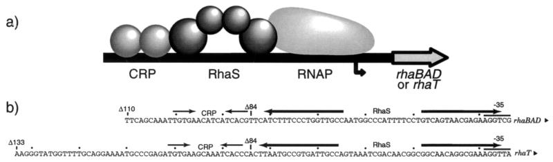
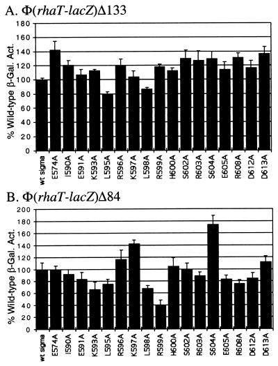
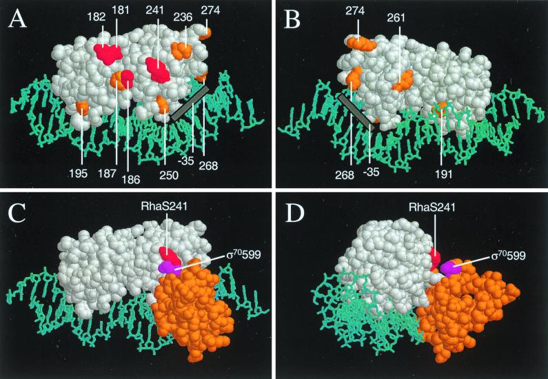
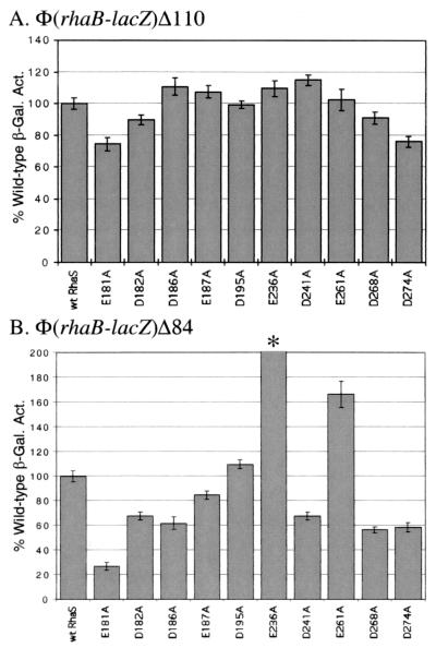
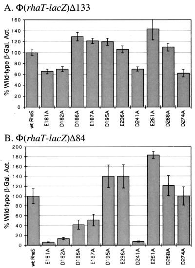
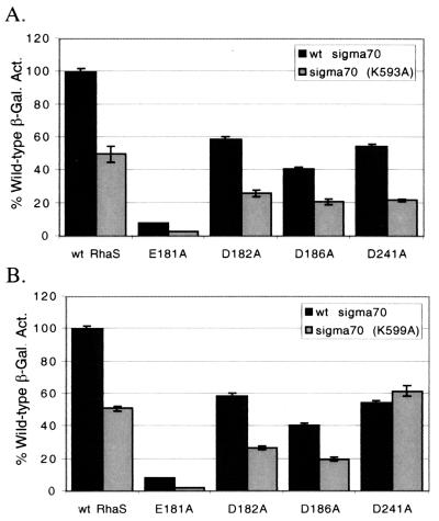
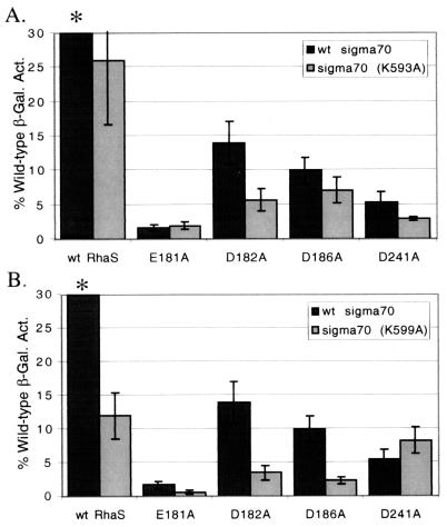
RhaS activates transcription of the Escherichia coli rhaBAD and rhaT operons in response to L-rhamnose and is a member of the AraC/XylS family of transcription activators. We wished to determine whether sigma(70) might be an activation target for RhaS. We found that sigma(70) K593 and R599 appear to be important for RhaS activation at both rhaBAD and rhaT, but only at truncated promoters lacking the binding site for the second activator, CRP. To determine whether these positively charged sigma(70) residues might contact RhaS, we constructed alanine substitutions at negatively charged residues in the C-terminal domain of RhaS. Substitutions at four RhaS residues, E181A, D182A, D186A, and D241A, were defective at both truncated promoters. Finally, we assayed combinations of the RhaS and sigma(70) substitutions and found that RhaS D241 and sigma(70) R599 met the criteria for interacting residues at both promoters. Molecular modeling suggests that sigma(70) R599 is located in very close proximity to RhaS D241; hence, this work provides the first evidence for a specific residue within an AraC/XylS family protein that may contact sigma(70). More than 50% of AraC/XylS family members have Asp or Glu at the position of RhaS D241, suggesting that this interaction with sigma(70) may be conserved.


Figures

FIG. 1
FIG. 1
(a) Schematic representation of the rhaBAD and rhaT promoter regions. RNA polymerase and the two activator proteins CRP and RhaS are shown bound to DNA in their respective positions. (b) DNA sequences of the rhaBAD and rhaT promoter regions, extending from the −35 regions to the most upstream endpoint of the promoter fusions used in this work. The positions of the RhaS-binding sites are shown by everted arrows, and the positions of the CRP-binding sites are shown by inverted arrows. The −35 regions of each promoter are marked, and the upstream endpoints of promoter fusions with lacZ are identified by a “Δ.”
FIG. 2
FIG. 2
Alanine substitutions in the ς70 subunit of E. coli RNA polymerase analyzed at a full-length fusion of the rhaBAD promoter with lacZ [Φ(rhaB-lacZ110] (A) and at a truncated fusion of the rhaBAD promoter with lacZ [Φ(rhaB-lacZ84] (B). In each case, a strain carrying the indicated translational fusion as a single-copy λ lysogen was transformed with a plasmid encoding either wild-type ς70 or a derivative with a single alanine substitution at the positions indicated. β-Galactosidase activity was measured from cultures grown in minimal medium with glycerol, l-rhamnose, and ampicillin. The x axis represents the ς70 derivative. The y axis represents the β-galactosidase specific activity for each ς70 derivative as a percentage of the activity for wild-type ς70. The wild-type activity in panel A was 402 Miller units, while the wild-type activity in panel B was 9 Miller units. Both were set to 100%.
FIG. 3
FIG. 3
Alanine substitutions in the ς70 subunit of E. coli RNA polymerase analyzed at a full-length fusion of the rhaT promoter with lacZ [Φ(rhaT-lacZ133] (A) and at a truncated fusion of the rhaT promoter with lacZ [Φ(rhaT-lacZ84] (B). In each case, a strain carrying the indicated translational fusion as a single-copy λ lysogen was transformed with a plasmid encoding either wild-type ς70 or a derivative with a single alanine substitution at the positions indicated. β-Galactosidase activity was measured in cultures grown in minimal medium with glycerol, l-rhamnose, and ampicillin. The x axis represents the ς70 derivative; the y axis represents the β-galactosidase specific activity for each ς70 derivative as a percentage of the activity for wild-type ς70. The wild-type activity in panel A was 2.5 Miller units; in panel B it was 0.079 Miller units. Both were set to 100%.
FIG. 4
FIG. 4
Model of the C-terminal domain of RhaS bound to DNA based on the crystal structure of a MarA-DNA complex (44). (A) “Front” view of RhaS C-terminal domain (white) in a space-filling model with the negatively charged residues highlighted and numbered. DNA is shown in a stick model and is colored cyan. RhaS residues (in red) were defective at both the rhaBAD and the rhaT promoters, while residues in orange were either not defective, were defective at only one promoter, or were not tested (D250 and D191). In this view the N-terminal subdomain of RhaS is on the left and the C-terminal subdomain is on the right. The approximate position of the −35 region of the promoter is shown as a gray bar. (B) Same as panel A, except rotated around the vertical axis by approximately 180° to give the “back” view (i.e., the N-terminal subdomain is on the right, and the C-terminal subdomain is on the left). (C) A model of the C-terminal region of ς70 (residues 550 to 613, orange, based on the DNA-binding domain of NarL) has been added to the RhaS C-terminal domain model. RhaS is in the same view as in panel A, but only the RhaS residue 241 is highlighted in red. The ς70 residue 599 is highlighted in violet. (D) Same as panel C, but rotated by somewhat less than 90° around the vertical axis. The modeling of ς70 onto the MarA-DNA complex was performed using the program Insight II, and panels A through D were drawn using RasMol version 2.6 for the Macintosh.
FIG. 5
FIG. 5
Alanine substitutions in RhaS analyzed at a full-length fusion of the rhaBAD promoter with lacZ [Φ(rhaB-lacZ110] (A) and at a truncated fusion of the rhaBAD promoter with lacZ [Φ(rhaB-lacZ84] (B). In each case, a strain carrying the indicated translational fusion as a single-copy λ lysogen was transformed with a plasmid encoding either wild-type RhaS or a derivative with a single alanine substitution at the positions indicated. β-Galactosidase activity was measured from cultures grown in minimal medium with glycerol, l-rhamnose, and ampicillin. The x axis represents the RhaS derivative. The y axis represents the β-galactosidase specific activity for each RhaS derivative as a percentage of the activity for wild-type RhaS. The wild-type activity in panel A was 453 Miller units for all of the assays except for E261A, where the wild-type activity was 204 Miller units, and in panel B it was 9.4 Miller units for all of the assays except for D241A, where the wild-type activity was 9.3 Miller units, and E261A, where wild-type activity was 3.9 Miller units. The activity in the case of E236A in panel B (marked with an asterisk) was 279%, but is drawn off the scale to avoid compression of the other values. The wild-type activity was set to 100%.
FIG. 6
FIG. 6
Alanine substitutions in RhaS analyzed at a full-length fusion of the rhaT promoter with lacZ [Φ(rhaT-lacZ133] (A) and a truncated fusion of the rhaT promoter with lacZ [Φ(rhaT-lacZ84] (B). In each case, a strain carrying the appropriate translational fusion as a single-copy λ lysogen was transformed with a plasmid encoding either wild-type RhaS or a derivative with a single alanine substitution at the positions indicated. β-Galactosidase activity was measured from cultures grown in minimal medium with glycerol, l-rhamnose, and ampicillin. The x axis represents the RhaS derivative. The y axis represents the β-galactosidase specific activity for each RhaS derivative as a percentage of the activity for wild-type RhaS. The wild-type activity in panel A was 1.38 Miller units for all of the assays except for with E261A, where the wild-type activity was 0.34 Miller units, and in panel B was 0.048 Miller units for all of the assays except for with E261A, where the wild-type activity was 0.027 Miller units. The wild-type activity was set to 100%.
FIG. 7
FIG. 7
Combinations of RhaS and ς70 alanine substitutions at Φ(rhaB-lacZ84. aRhaS substitutions were tested in combination with either ς70 K593A (A) or ς70 R599A (B) at the Φ(rhaB-lacZ84 fusion. The β-galactosidase specific activity for each combination is represented as a percentage of the activity found for the combination of wild-type RhaS and wild-type ς70, which was 9.1 Miller units and was set to 100% for both graphs.
FIG. 8
FIG. 8
Combinations of RhaS and ς70 alanine substitutions at Φ(rhaT-lacZ84. aRhaS substitutions were tested in combination with either ς70 K593A (A) or ς70 R599A (B) at the Φ(rhaT-lacZ84 fusion. The β-galactosidase specific activity for each combination is represented as the percentage of the activity found for the combination of wild-type RhaS and wild-type ς70, which was 0.18 Miller units and was set to 100% for both graphs.

References

    1. Akimuru H, Sakumi K, Yoshikai T, Anai M, Sekiguchi M. Positive and negative regulation of transcription by a cleavage product of Ada protein. J Mol Biol. 1990;216:261–273. - PubMed
    1. Backman K, Chen Y-M, Magasanik B. Physical and genetic characterization of the gln A-glnG region of the Escherichia coli chromosome. Proc Natl Acad Sci USA. 1981;78:3743–3747. - PMC - PubMed
    1. Baikalov I, Schroder I, Kaczor-Grzeskowiak M, Grzeskowaik K, Gunsalus R P, Dickerson R E. Structure of the Escherichia coli response regulator NarL. Biochemistry. 1996;35:11053–11061. - PubMed
    1. Bhende P M, Egan S M. Amino acid-DNA contacts by RhaS: an AraC family transcription activator. J Bacteriol. 1999;181:5185–5192. - PMC - PubMed
    1. Bourgerie S J, Michan C M, Thomas M S, Busby S J W, Hyde E I. DNA binding and DNA bending by the MelR transcription activator protein from Escherichia coli. Nucleic Acids Res. 1997;25:1685–1693. - PMC - PubMed

Publication types

MeSH terms