. 2000 Sep;47(3):415-22.
doi: 10.1136/gut.47.3.415.

Comparable expression of matrix metalloproteinases 1 and 2 in pouchitis and ulcerative colitis

Affiliations

Comparable expression of matrix metalloproteinases 1 and 2 in pouchitis and ulcerative colitis

A Stallmach et al. Gut. 2000 Sep.

Abstract

Background and aims: Matrix metalloproteinases (MMPs) are implicated in the tissue destruction associated with inflammatory diseases. Proctocolectomy with ileo-anal pouch (IAP) anastomosis is associated with pouchitis, particularly in patients with ulcerative colitis (UC). The aim of this study was to quantify MMP-1 and MMP-2 in inflamed and uninflamed pouches of patients with UC compared with those with active UC. IAP patients with familial adenomatous polyposis (FAP) served as controls.

Methods: Biopsies were taken from 33 patients with IAP (UC, n=25; FAP, n=8) and from 10 UC patients. MMP-1 and MMP-2 were quantified using sandwich enzyme linked immunosorbent assays. In addition, northern and western blotting and in situ hybridisation experiments were performed.

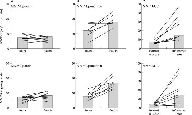
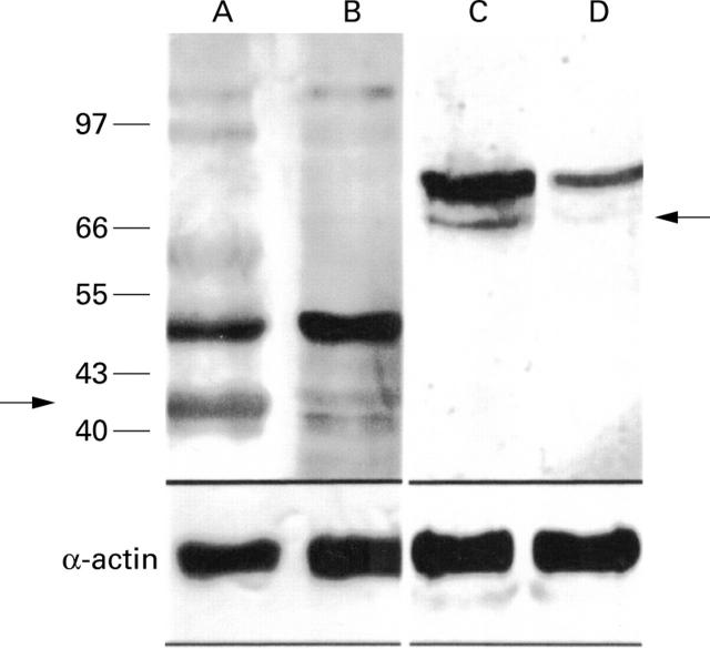
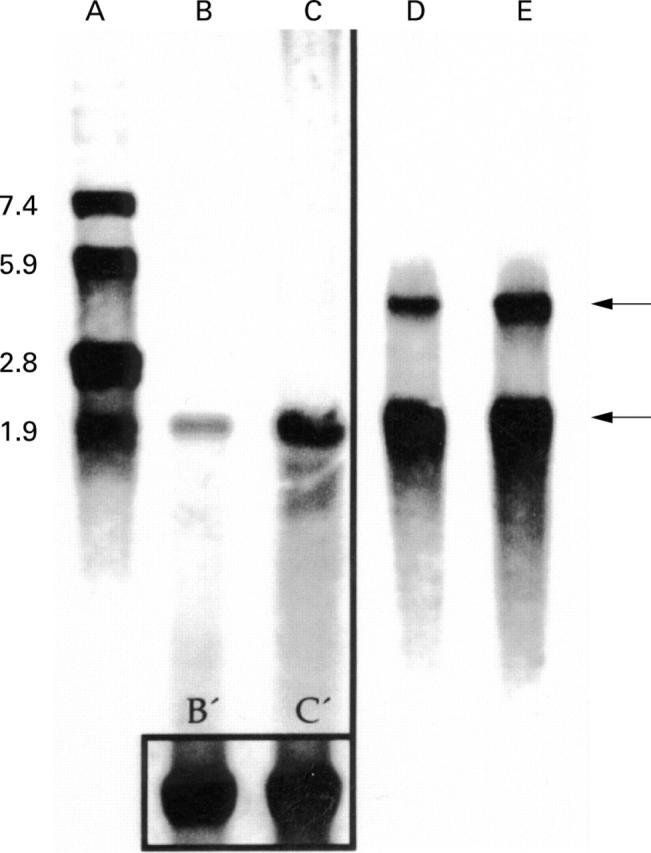
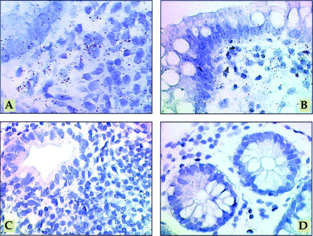
Results: In pouchitis (n=11), MMP-1 and MMP-2 concentrations were increased compared with uninflamed pouches of patients with UC (n=14) or FAP (n=8) (MMP-1 17.7 ng/mg protein v 7.8 (UC) v 7.6 (FAP), p</=0.05; MMP-2 16.4 v 9.5 (UC) v 6.3 (FAP), p</=0.05). Western and northern blots revealed increased MMP-1 and MMP-2 protein and transcript concentrations in inflamed pouches. Mesenchymal cells were identified as major producers of MMP-1 and MMP-2 in pouchitis. A similar increase in MMPs was observed in tissues of patients with active UC.

Conclusions: Our results support the hypothesis that MMPs are involved in mucosal destruction and crypt hyperplasia, as seen in pouchitis.


Figures

Figure 1
Figure 1
MMP-1 and MMP-2 concentrations in uninflamed pouches, in pouchitis, and in active ulcerative colitis (UC). Mucosal biopsies were taken from ileo-anal pouches and the adjacent ileum from individual patients without (A) (n=10) or with pouchitis (B) (n=7) and from patients with active UC (C) (n = 10). For technical reasons it was not possible to take ileal biopsies in one patient with pouchitis and four patients with uninflamed pouches. MMP-1 and MMP-2 concentrations were determined as described in material and methods. Columns represent the median of all patients; lines connect intraindividual MMP-1 and MMP-2 concentrations.
Figure 2
Figure 2
Concentrations of MMP-1 and MMP-2 after treatment of pouchitis. Mucosal biopsies were taken from ileo-anal pouches from individual patients (n=7) with pouchitis before and after treatment with metronidazole for two weeks, and analysed for MMP-1 and MMP-2 concentrations. Lines connect the follow up values for MMP concentrations before and after normalisation of clinical and endoscopic signs of inflammation; bars represent median of all patients. In one patient (*), endoscopic follow up after treatment did not show a significant improvement in mucosal inflammation. However, clinical and histological parameters improved.
Figure 3
Figure 3
Western blot analysis of MMP-1 and MMP-2 expression. Western blotting revealed increased concentrations of MMP proenzymes in pouchitis. Lanes A and C, pouchitis; lanes B and D, uninflamed pouch. Lanes A and B, MMP-1; lanes C and D, MMP-2 (arrows indicate the 42 kDa MMP-1 and the 66 kDa MMP-2). The lower part of the figure shows results of western blot analysis using an α-actin specific antibody to demonstrate equal protein load on each lane.
Figure 4
Figure 4
Northern blot analysis of MMP-1- and MMP-2 in mucosal biopsies from patients with pouchitis. Northern blot analysis of MMP-1 (lanes B, C) and MMP-2 (lanes D and E, upper arrow 3.1 kB) and β-actin RNA (lower arrow 1.7 kB). The box in the lower part of lanes B and C represents β-actin RNA of B and C. Lane A represents the RNA bp standard. mRNA was extracted from biopsies from the adjacent uninflamed ileum serving as an internal control (lanes B and D) and inflamed pouch (lanes C and E). A representative example of one of five patients is shown.
Figure 5
Figure 5
Mesenchymal cells are major producers of MMP-1 and MMP-2 in pouchitis. In situ hybridisation with [35S] labelled MMP-1 antisense RNA probe (A) or [35S] labelled MMP-2 antisense RNA probe (B) in pouchitis. In inflamed pouches a large number of strongly labelled cells is visible in the lamina propria, mainly directly underneath the epithelium (A, B). [35S] labelled MMP-1 sense RNA probe (C) in pouchitis, demonstrating the background signal. In situ hybridisation with [35S] labelled MMP-2 antisense RNA probe (D) on ileum tissue with mild inflammation. In the uninflamed ileum grains are localised over mesenchymal cells of the lamina propria, predominantly underneath the epithelium. However, transcript levels were near the threshold of detection in most cases. Exposure time in all sections was 28 days. Original magnification ×600.

References

    1. Br Med J. 1978 Jul 8;2(6130):85-8 - PubMed
    1. Matrix Biol. 1999 Aug;18(4):373-80 - PubMed
    1. World J Surg. 1987 Dec;11(6):727-34 - PubMed
    1. Nature. 1989 Feb 16;337(6208):661-3 - PubMed
    1. FASEB J. 1991 May;5(8):2145-54 - PubMed

Publication types

Substances