

Expression of interleukin 8 (IL-8) and substance P in human chronic pancreatitis
- PMID: 10940282
- PMCID: PMC1728055
- DOI: 10.1136/gut.47.3.423
Expression of interleukin 8 (IL-8) and substance P in human chronic pancreatitis
Abstract
Background: Changes in substance P content and a relationship between the degree of perineural inflammation and pain has been demonstrated in chronic pancreatitis. Whether a relationship exists between neural alteration and pancreatic inflammation (neurogenic inflammation) is not known.
Aims: In the present study we evaluated gene expression of preprotachykinin A (PPT-A), the gene encoding substance P, and interleukin 8, a proinflammatory and hyperalgesic mediator whose release is co-regulated by substance P.
Patients: Pancreatic tissue specimens obtained from 21 patients (16 male, five female) with chronic pancreatitis and 18 healthy organ donors (nine male, nine female) were analysed.
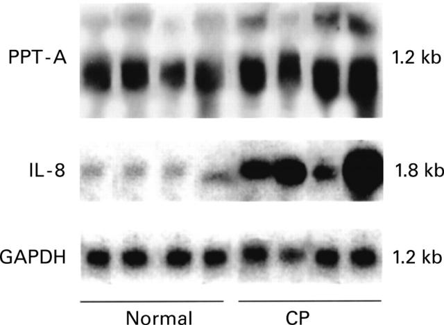
Methods: Gene expression of PPT-A and interleukin 8 was studied by northern blot analysis. Respective proteins were localised using immunohistochemistry.
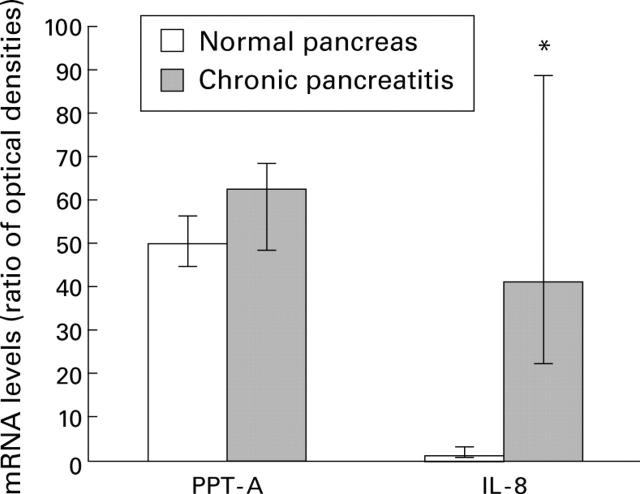
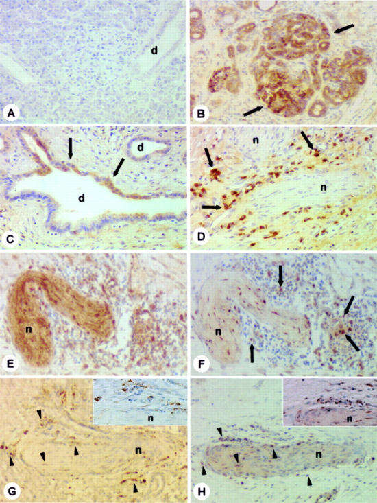
Results: Northern blot analysis showed that PTT-A mRNA expression levels were present at comparable levels in normal and chronic pancreatitis tissue samples. In contrast, interleukin 8 mRNA was expressed at very low levels in normal controls but was increased 41-fold (p<0. 001) in chronic pancreatitis tissue samples. Using immunohistochemistry, interleukin 8 protein was localised mainly in immune cells often found around enlarged pancreatic nerves. In addition, in chronic pancreatitis, intense interleukin 8 immunostaining was present in metaplastic ductal cells of the atrophic pancreatic parenchyma. In chronic pancreatitis samples there was a positive relationship between interleukin 8 mRNA levels and the presence of ductal metaplasia (r=0.795; p<0.001) and the inflammation score (r=0.713; p<0.001).
Conclusions: Our data indicate that in chronic pancreatitis, the increase in substance P in enlarged pancreatic nerves is not caused by enhanced intrapancreatic PTT-A mRNA expression, suggesting that the location of substance P synthesis is outside of the pancreas. In addition, localisation of interleukin 8 positive immune cells around pancreatic nerves further supports the existence of neuroimmune interactions as a pathophysiological mechanism in chronic pancreatitis.
Figures
References
MeSH terms
Substances
LinkOut - more resources
Full Text Sources
Other Literature Sources
Medical