. 2000 Aug 15;19(16):4228-36.
doi: 10.1093/emboj/19.16.4228.

Crystal structure of the matrix protein VP40 from Ebola virus

Affiliations

Crystal structure of the matrix protein VP40 from Ebola virus

A Dessen et al. EMBO J. .

Abstract

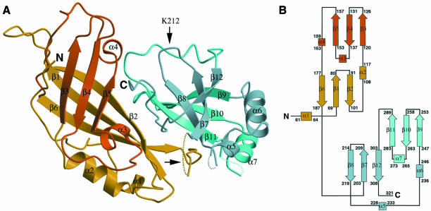
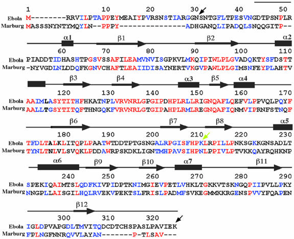
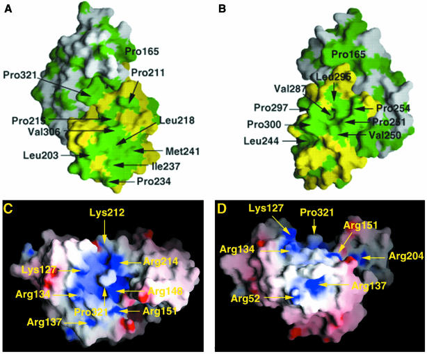
Ebola virus maturation occurs at the plasma membrane of infected cells and involves the clustering of the viral matrix protein VP40 at the assembly site as well as its interaction with the lipid bilayer. Here we report the X-ray crystal structure of VP40 from Ebola virus at 2.0 A resolution. The crystal structure reveals that Ebola virus VP40 is topologically distinct from all other known viral matrix proteins, consisting of two domains with unique folds, connected by a flexible linker. The C-terminal domain, which is absolutely required for membrane binding, contains large hydrophobic patches that may be involved in the interaction with lipid bilayers. Likewise, a highly basic region is shared between the two domains. The crystal structure reveals how the molecule may be able to switch from a monomeric conformation to a hexameric form, as observed in vitro. Its implications for the assembly process are discussed.


Figures

None
Fig. 1. Stereo view of the experimental map generated with MAD phases obtained from six selenium sites identified by SOLVE and subsequently solvent flattened. The map is contoured at the 1σ level, and focuses on a conserved loop region in domain 2 connecting β-strands 7 and 8.
None
Fig. 2. (A) Ribbon diagram of Ebola VP40. Two domains are shown in different colours. Secondary structure elements coloured yellow and dark blue lie on a plane below those coloured orange and light blue. Flexible loop regions, which were not traceable in the experimental maps, including the interdomain connecting loop (horizontal arrow) are drawn as dashed lines. The trypsin cleavage site, after Lys212, is highlighted by a vertical arrow. Trypsinization at this site causes complete disengagement of the C-terminal domain from the N-terminus, and subsequent hexamerization of the N-terminal domain. (B) Topology diagram of the two domains of Ebola virus VP40. β-strands are represented as arrows, while α-helices are rectangles. Short helical turns are represented as small squares. The domains display structural resemblance.
None
Fig. 3. Sequence alignment of VP40 from Ebola virus (strain Zaire, DDBJ/EMBL/GenBank accession No. AF086833) and from Marburg virus (strain Popp, DDBJ/EMBL/GenBank accession No. Z29337). Identical residues are shown in red letters and conservative substitutions in blue. The sequence present in the structure is shown schematically with the assignment of secondary structure elements (based on the structure of Ebola virus VP40). The black arrows at positions 31 and 326 indicate the first and last residues present in the construct used for crystallization. The yellow arrow at position 212 indicates the position of the trypsin cleavage site that induces hexamerization of VP40 in solution in vitro (Ruigrok et al., 2000b). The numbering is according to the Ebola virus VP40 sequence.
None
Fig. 4. Interdomain interactions. (A) Close up of polar interactions between the N- and C-terminal domains. Residues involved in salt bridges and hydrogen bonds are shown. For clarity, the connection between residue 307 and 310 is shown as a grey dashed line. The loop connecting both domains is indicated with an arrow. (B) Surface representation of the N-terminal domain (residues 44–194) and (C) of the C-terminal domain (residues 201–321). Hydrophobic residues lining the interface on the N- and C-terminal domains are shown in green.
None
Fig. 5. Distribution of hydrophobic and electrostatic surfaces. (A and B) Surface representation of VP40 in two orientations, rotated by 90° with respect to each other. The N-terminal domain is shown in white and the C-terminal domain in yellow. Hydrophobic residues are depicted in green. (C and D) Charge distribution of Ebola virus VP40 (two orientations, rotated by 90°). Surface potential representation of VP40 with regions where the electrostatic potential is less than –10 kBT are red, while those more than +10 kBT are blue (kB, Boltzmann constant; T, absolute temperature). A basic patch is clearly visible in the central region of the molecule and encompasses residues from both domains.

Similar articles

Cited by

References

    1. Becker S., Spiess,M. and Klenk,H.D. (1995) The asialoglycoprotein receptor is a potential liver-specific receptor for Marburg virus. J. Gen. Virol., 76, 393–399. - PubMed
    1. Brünger A.T. et al. (1998) Crystallographic and NMR system (CNS): a new software system for macromolecular structure determination. Acta Crystallogr. D, 54, 905–921. - PubMed
    1. Bukreyev A.A., Volchkov,V.E., Blinov,V.M. and Netesov,S.V. (1993) The Vp35 and VP40 proteins of filoviruses. Homology between Marburg and Ebola viruses. FEBS Lett., 322, 41–46. - PubMed
    1. Christensen A.M., Massiah,M.A., Turner,B.G., Sundquist,W.I. and Summers,M.F. (1996) Three-dimensional structure of the HTLV-II matrix protein and comparative analysis of matrix proteins from different classes of pathogenic human retroviruses. J. Mol. Biol., 264, 1117–1131. - PubMed
    1. Collaborative Computational Project Number 4 (1994) The CCP4 suite: programs for protein crystallography. Acta Crystallogr. D, 50, 760–776. - PubMed

Publication types

Associated data