. 2000 Aug 15;19(16):4310-22.
doi: 10.1093/emboj/19.16.4310.

Negative regulation of cytochrome c-mediated oligomerization of Apaf-1 and activation of procaspase-9 by heat shock protein 90

Affiliations

Negative regulation of cytochrome c-mediated oligomerization of Apaf-1 and activation of procaspase-9 by heat shock protein 90

P Pandey et al. EMBO J. .

Abstract

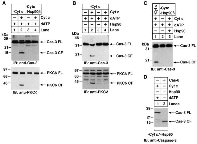
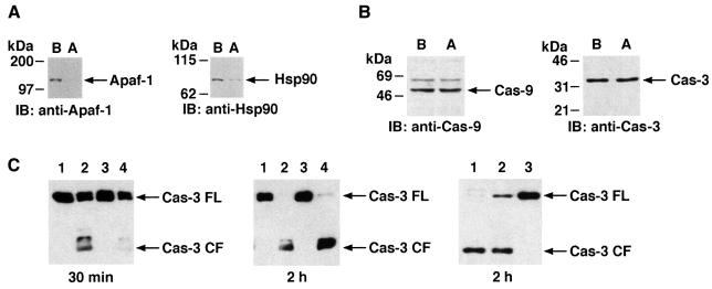
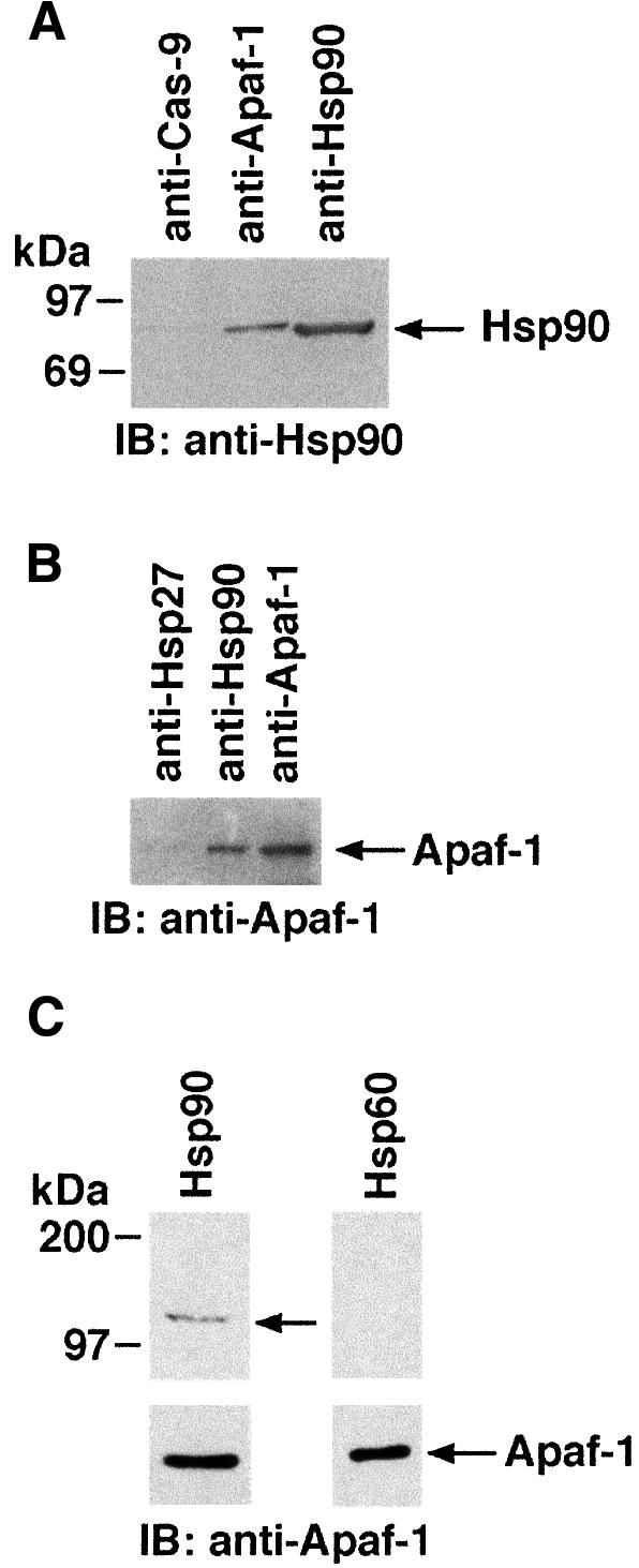
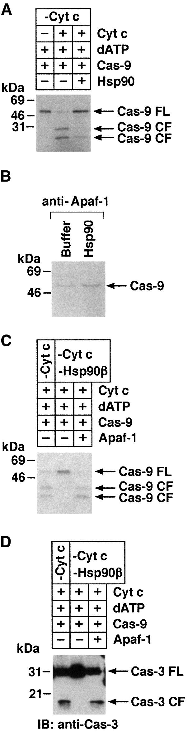
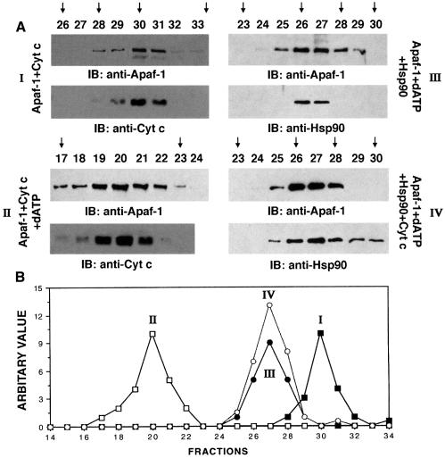
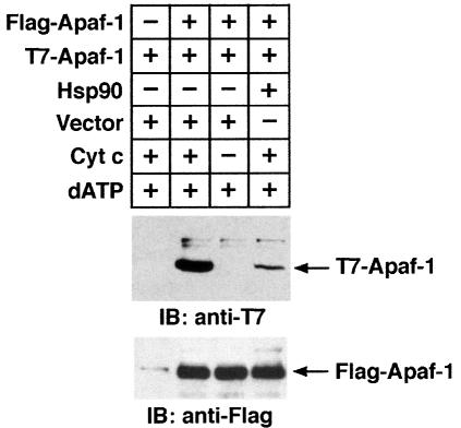
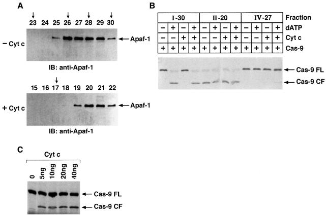
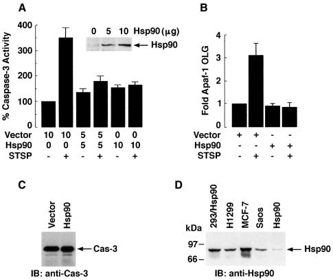
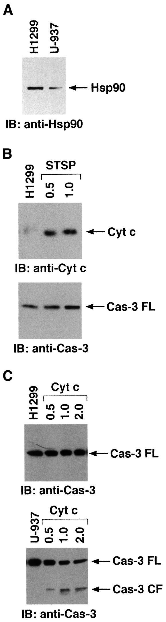
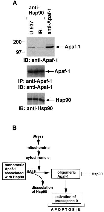
The release of cytochrome c from mitochondria results in the formation of an Apaf-1-caspase-9 apoptosome and induces the apoptotic protease cascade by activation of procaspase-3. The present studies demonstrate that heat shock protein 90 (Hsp90) forms a cytosolic complex with Apaf-1 and thereby inhibits the formation of the active complex. Immunodepletion of Hsp90 depletes Apaf-1 and thereby inhibits cytochrome c-mediated activation of caspase-9. Addition of purified Apaf-1 to Hsp90-depleted cytosolic extracts restores cytochrome c-mediated activation of procaspase-9. We also show that Hsp90 inhibits cytochrome c-mediated oligomerization of Apaf-1 and thereby activation of procaspase-9. Furthermore, treatment of cells with diverse DNA-damaging agents dissociates the Hsp90-Apaf-1 complex and relieves the inhibition of procaspase-9 activation. These findings provide the first evidence for a negative cytosolic regulator of cytochrome c-dependent apoptosis and for involvement of a chaperone in the caspase cascade.


Figures

None
Fig. 1. Detection of cytosolic proteins that associate with cyt c. (A) U-937 cells were disrupted in a Dounce homogenizer and the S-100 extract was applied to a cyt c bound to CNBr–sepharose beads column. After washing, elution was performed with a linear NaCl gradient (0–600 mM). The eluted proteins were separated by SDS–PAGE and analyzed by silver staining. (B) Following elution from cyt c, adsorbed proteins were separated by SDS–PAGE and then transferred to PVDF membranes. After localization of a 90 kDa protein by Ponceau staining, tryptic digestion and amino acid sequence of the resulting peptides was performed (Worcester Foundation for Biomedical Research, Shrewsbury, MA). Sequence analysis of the 90 kDa protein is compared with that of the Hsp90β protein. (C) Eluted fractions from the cyt c–CNBr–sepharose column were separated by 10% SDS–PAGE and analyzed by immunoblotting with anti-Hsp90β antibody.
None
Fig. 2. (A) Lysates from U-937 cells were subjected to immunoprecipitation with anti-Hsp90β or anti-cyt c. The resulting protein precipitates were analyzed by immunoblotting with anti-Hsp90β. (B) Lysates from U-937 cells were subjected to immunoprecipitation with anti-Hsp90β or anti-cyt c and the precipit ates were analyzed by immunoblotting with anti-cyt c.
None
Fig. 3. Depletion of Hsp90β abrogates cyt c-mediated activation of caspase-3 in an in vitro cell-free system. (A) U-937 S-100 extracts were immunodepleted of cyt c (lanes 1–4) or Hsp90β (lanes 3 and 4). Aliquots (50 µg) were then incubated with buffer (lanes 1 and 3) or purified cyt c (1 µg, lanes 2 and 4) in the presence of 1 mM dATP in a final volume of 30 µl. After incubation at room temperature for 1 h, the proteins were separated by 10% SDS–PAGE and analyzed by immunoblotting with anti-caspase-3 (upper panel) or anti-PKCδ (lower panel). FL, full length; CF, cleaved fragment. (B) U-937 S-100 extracts were immunodepleted of cyt c (lanes 1–4). Aliquots were then incubated with buffer (lane 1), purified cyt c (lanes 2 and 4) or purified Hsp90 (1 µg; lanes 3 and 4) and analyzed by immunoblotting with anti-caspase-3 (upper panel) or anti-PKCδ (lower panel). (C) U-937 cell S-100 extracts were immunodepleted of cyt c (lanes 1–3) or Hsp90β (lanes 2 and 3). Aliquots (50 µg) were then incubated with purified cyt c (1 µg, lanes 1 and 3) or with Hsp90 (lane 3) in the presence of 1 mM dATP in a final volume of 30 µl. After incubation at room temperature for 1 h, the proteins were separated by 10% SDS–PAGE and analyzed by immunoblotting with anti-caspase-3 or anti-PKCδ. (D) U-937 cytosolic lysates were immunodepleted of cyt c and Hsp90β. Aliquots (50 µg) were then incubated with cyt c (1 µg) or purified recombinant caspase-8 in the presence of 1 mM dATP in a final volume of 30 µl. After incubation for 30 min at 30°C, the proteins were separated by SDS–PAGE and analyzed by immunoblotting with anti-caspase-3.
None
Fig. 4. Hsp90 associates with Apaf-1. (A) U-937 cell S-100 extracts were immunodepleted of Hsp90. Lysates from lane B (before immunodepletion) and lane A (after immunodepletion) were subjected to SDS–PAGE and analyzed by immunoblotting with anti-Apaf-1 (left panel) or anti-Hsp90 (right panel). (B) Lysates from before and after immunodepletion were also subjected to SDS–PAGE and analyzed by immunoblotting with anti-caspase-9 (left panel) or anti-caspase-3 (right panel). (C) S-100 extracts were immunodepleted of cyt c (lanes 1–4) and Hsp90 (lanes 3 and 4). Aliquots (50 µg) were then incubated with buffer (lanes 1 and 3) or purified cyt c (1 µg, lanes 2 and 4) in the presence of 1 mM dATP in a final volume of 30 µl. After incubation at room temperature for 30 min (left panel) or 2 h (middle panel), the proteins were separated by 10% SDS–PAGE and analyzed by immunoblotting with anti-caspase-3. Right panel: S-100 extracts were immunodepleted of cyt c (lanes 1–3) and Hsp90β (lanes 2 and 3). Aliquots (50 µg) were then incubated with purified cyt c/dATP (lanes 1–3) and Hsp90 (lane 3) at room temperature for 2 h. Proteins were separated by 10% SDS–PAGE and analyzed by immunoblotting with anti-caspase-3.
None
Fig. 5. (A) Lysates from U-937 cells were subjected to immunoprecipitation with anti-Hsp90, anti-caspase-9 or anti-Apaf-1. The resulting protein precipitates were then analyzed by immunoblotting with anti-Hsp90. (B) U-937 lysates were subjected to immunoprecipitation with anti-Hsp90, anti-Hsp27 or anti-Apaf-1 and analyzed by immunoblotting with anti-Apaf-1. (C) Column-purified Apaf-1 protein was resolved by SDS–PAGE and transferred to nitocellulose filters. The filters were incubated with either Hsp60 or Hsp90 protein. The filters were then analyzed by immunoblotting with anti-Hsp60 or anti-Hsp90 antibody (top panels). The same filters were also analyzed by immunoblotting with anti-Apaf-1 (bottom panels).
None
Fig. 6. (A) U-937 cytosolic lysates were immunodepleted of cyt c (lanes 1–3). Aliquots (50 µg) were then incubated with buffer (lane 1), cyt c (lanes 2 and 3) or Hsp90 (lane 3). In vitro-translated 35S-labeled caspase-9 (2 µl) was incubated in a final volume of 30 µl for 1 h at 30°C. After incubation, samples were separated by SDS–PAGE and analyzed by autoradiography. (B) Column-purified Apaf-1 was incubated with in vitro-translated 35S-labeled caspase-9 and cyt c/dATP in the presence of Hsp90 or buffer. After incubation, anti-Apaf-1 immuno precipitates were analyzed by autoradiography. (C and D) U-937 cell cytosolic lysates were immunodepleted of cyt c (lanes 1–3) or cyt c/Hsp90 (lanes 2 and 3). Aliquots (50 µg) were then incubated with cyt c/dATP (lanes 1–3), in vitro-translated 35S-labeled caspase-9 (D, lanes 1–3) in the presence (lane 3) or absence (lanes 1 and 2) of purified Apaf-1 L-WD13 in a final volume of 30 µl. After incubation at 30°C for 1 h, the proteins were separated by SDS–PAGE and analyzed by immunoblotting with anti-caspase-3 (C) or autoradiography (D).
None
Fig. 7. Hsp90 inhibits cyt c/dATP-mediated oligomerization of Apaf-1. (A) Purified Apaf-1 L-WD13 was incubated with cyt c (I), cyt c + dATP (II), dATP + Hsp90 (III) or dATP + cyt c + Hsp90 (IV). After incubation, the reaction mixtures were applied to Superose-6 FPLC column at a flow rate of 0.2 ml/min. Aliquots (45 µl) of the column fractions in each case were separated by SDS–PAGE and immunoblotted with anti-Apaf-1, anti-cyt c or anti-Hsp90. The calibration protein standards and their positions on the Superose-6 FPLC column are indicated by vertical arrows: fraction 17, 2 MDa; fraction 23, 669 kDa; fraction 26, 440 kDa, fraction 28, 232 kDa; fraction 30, 158 kDa; fraction 34, 66 kDa. (B) An elution profile of Apaf-1 L-WD13 under different conditions (I–IV) is shown.
None
Fig. 8. Hsp90 inhibits oligomerization of Apaf-1. 293T cells were transiently transfected with Flag-Apaf-1 (lanes 2–4) and T7-Apaf-1 (lanes 1–4) with (lane 4) or without (lanes 1–3) Hsp90. Cytosolic lysates were incubated with (lanes 1, 2 and 4) or without (lane 3) cyt c and subjected to immunoprecipitation with anti-Flag. The immuno precipitates were then analyzed by immunoblotting with anti-T7 (upper panel). Anti-Flag immunoprecipitates were also analyzed by immunoblotting with anti-Flag (lower panel).
None
Fig. 9. Hsp90 inhibits Apaf-1-mediated activation of caspase-9. (A) Fraction IV-26 and IV-27 from Apaf-1 + dATP + Hsp90 column eluate were pooled, concentrated and incubated at room temperature in the absence (–cyt c) or presence (+cyt c) of cyt c for 1 h. Following incubation, samples were applied to a Superose-6 FPLC column at a flow rate of 0.2 ml/min. Aliquots (45 µl) of the column fractions in each case were separated by SDS–PAGE and immunoblotted with anti-Apaf-1. (B35S-labeled caspase-9 was incubated with peak fractions (I-30, II-20 or IV-27) in the presence or absence of cyt c/dATP. Following incubation, samples were then analyzed by SDS–PAGE and autoradiography. Full-length caspase-9 (FL) and the p35 fragment of mature caspase-9 (CF) are shown. (C) Peak fraction from Apaf-1 + dATP + Hsp90 column eluate was incubated with 35S-labeled caspase-9 in the absence (lane 1) or presence (lanes 2–5) of increasing concentrations of cyt c. Following incubation, samples were analyzed by SDS–PAGE and autoradiography.
None
Fig. 10. Hsp90 inhibits oligomerization of Apaf-1 and activation of caspase-3 in response to stress. (A) L929 cells were transfected with empty vector or 5 and 10 µg of vector expressing Hsp90. Cells were also cotransfected with pEGFP. Following transfection, GFP-positive cells were selected by FACS, stabilized in culture for 24 h and treated with 1 µg/ml STSP for 18 h. Total lysates from untreated and STSP-treated cells were analyzed for caspase-3 activity by measuring cleavage of an exogenous peptide substrate (DEVD-pNA). Results are expressed as percentage caspase-3 activity (mean ± SD) (normalized to untreated cells expressing vector) of four independent experiments. Total cell lysates were also analyzed by immunoblotting with anti-Hsp90 (insert). (B) 293T cells were cotransfected with T7-Apaf-1, Flag-Apaf-1 and empty vector or Hsp90. Following transfection, cells were treated with 1 µM STSP and harvested after 18 h. Total cell lysates were subjected to immunoprecipitation with anti-T7 and analyzed by immunoblotting with anti-Flag. Results are expressed as fold-Apaf-1 oligomerization (mean + SD) of three independent experiments. (C) 293T cells were transiently transfected with empty vector or Hsp90. Total cell lysates were analyzed by immunoblotting with anti-caspase-3. (D) 293T cells were transiently transfected with Hsp90. Total lysates from transfected 293T cells and lysates of H1299, MCF-7 and SAOS cells were analyzed by immunoblotting with anti-Hsp90. Hsp90-immunodepleted S-100 extract of U-937 cells was incubated with 1 µg Hsp90 and analyzed by immunoblotting with anti-Hsp90.
None
Fig. 11. (A) Total cell lysates from H1299 and U-937 cells were analyzed by immunoblotting with anti-Hsp90. (B) H1299 cells were treated with 0.5 or 1 µM STSP and harvested after 18 h. S-100 cytosolic extracts were prepared and analyzed by immunoblotting with anti-cyt c (upper panel) or anti-caspase-3 (lower panel). (C) S-100 cytosolic extracts from untreated H1299 (upper panel) or U-937 (lower panel) cells were incubated with 0, 0.25, 0.5 or 1 µg of purified cyt c in the presence of 1 mM dATP for 30 min at room temperature. Following incubation, proteins were separated by SDS–PAGE and analyzed by immunoblotting with anti-caspase-3.
None
Fig. 12. (A) U-937 cells were exposed to 20 Gy IR and harvested at 6 h. Cytosolic lysates were immunoprecipitated with anti-Hsp90 and analyzed by immunoblotting with anti-Apaf-1 (upper panel). Anti-Apaf-1 immunoprecipitates were analyzed separately by immunoblotting with anti-Apaf-1 (middle panel). Total cell lysates were also analyzed by immunoblotting with anti-Hsp90 (lower panel). (B) Schematic representation of the role of Hsp90 in cyt c/dATP-dependent oligomerization and activation of Apaf-1.

Similar articles

Cited by

References

    1. Adrain C., Slee,E., Harte,M. and Martin,S. (1999) Regulation of apoptotic protease activating factor-1 oligomerization and apoptosis by the WD-40 repeat region. J. Biol. Chem., 274, 20855–20860. - PubMed
    1. Ali A., Krone,P., Pearson,D. and Heikkila,J. (1996) Evaluation of stress-inducible hsp90 gene expression as a potential molecular biomarker in Xenopus laevis.Cell Stress Chaperones, 1, 62–69. - PMC - PubMed
    1. Alnemri E.S., Livingston,D.J., Nicholson,D.W., Salvesen,G., Thornberry,N.A., Wong,W.W. and Yuan,J. (1996) Human ICE/CED-3 protease nomenclature. Cell, 87, 171. - PubMed
    1. Benedict M., Hu,Y., Inohara,N. and Nunez,G. (2000) Expression and functional analysis of Apaf-1 isoforms. Extra WD-40 repeat is required for cytochrome c binding and regulated activation of procaspase-9. J. Biol. Chem., 275, 8461–8468. - PubMed
    1. Bertram J., Palfner,K., Hiddemann,W. and Kneba,M. (1996) Increase of P-glycoprotein-mediated drug resistance by hsp90 β. Anti-Cancer Drugs, 7, 838–845. - PubMed

Publication types

MeSH terms