. 2000 Aug 15;527 Pt 1(Pt 1):131-7.
doi: 10.1111/j.1469-7793.2000.t01-2-00131.x.

Twitch and tetanic force responses and longitudinal propagation of action potentials in skinned skeletal muscle fibres of the rat

Affiliations

Twitch and tetanic force responses and longitudinal propagation of action potentials in skinned skeletal muscle fibres of the rat

G S Posterino et al. J Physiol. .

Abstract

1. Transverse electrical field stimulation (50 V cm-1, 2 ms duration) of mechanically skinned skeletal muscle fibres of the rat elicited twitch and tetanic force responses (36 +/- 4 and 83 +/- 4 % of maximum Ca2+-activated force, respectively; n = 23) closely resembling those in intact fibres. The responses were steeply dependent on the field strength and were eliminated by inclusion of 10 microM tetrodotoxin (TTX) in the (sealed) transverse tubular (T-) system of the skinned fibres and by chronic depolarisation of the T-system. 2. Spontaneous twitch-like activity occurred sporadically in many fibres, producing near maximal force in some instances (mean time to peak: 190 +/- 40 ms; n = 4). Such responses propagated as a wave of contraction longitudinally along the fibre at a velocity of 13 +/- 3 mm s-1 (n = 7). These spontaneous contractions were also inhibited by inclusion of TTX in the T-system and by chronic depolarisation. 3. We examined whether the T-tubular network was interconnected longitudinally using fibre segments that were skinned for only approximately 2/3 of their length, leaving the remainder of each segment intact with its T-system open to the bathing solution. After such fibres were exposed to TTX (60 microM), the adjacent skinned region (with its T-system not open to the solution) became unresponsive to subsequent electrical stimulation in approximately 50 % of cases (7/15), indicating that TTX was able to diffuse longitudinally inside the fibre via the tubular network over hundreds of sarcomeres. 4. These experiments show that excitation-contraction coupling in mammalian muscle fibres involves action potential propagation both transversally and longitudinally within the tubular system. Longitudinal propagation of action potentials inside skeletal muscle fibres is likely to be an important safety mechanism for reducing conduction failure during fatigue and explains why, in developing skeletal muscle, the T-system first develops as an internal longitudinal network.


Figures

Figure 1
Figure 1. Twitch and tetanic force responses elicited in a ‘skinned’ skeletal muscle fibre devoid of surface membrane
A, schematic representation of the skinning of a single muscle fibre by rolling back the surface membrane (sarcolemma) with a pair of forceps, forming a ‘cuff’. The transverse tubular system (T) seals off to form a closed compartment after skinning. S, sarcolemma; SR, sarcoplasmic reticulum; Z, Z-line; C, contractile apparatus; F, forceps. B, a skinned segment of a rat EDL muscle fibre was mounted on a force transducer and bathed in a solution mimicking the normal cytoplasmic environment (high [K+]), re-establishing the normal resting membrane potential in the sealed T-system. From top to bottom: a single twitch, an unfused tetanus and a fused tetanus (50 Hz) elicited by applying 50 V cm−1 field stimulation (2 ms duration) between two platinum wire electrodes running parallel to the long axis of the fibre. Individual arrows indicate single stimuli and the filled bar indicates a continuous 50 Hz stimulus. C, force response in the same skinned segment elicited when the sealed T-system was depolarised by replacing the K+-based bathing solution with a Na+-based solution, and assessment of maximum force by direct activation of the contractile apparatus with a heavily buffered solution with 20 μm free Ca2+ (Max Act). Fibre segment length (L), 1.2 mm; diameter (D), 50 μm.
Figure 2
Figure 2. Twitches in a skinned fibre are elicited by the generation of an action potential in the sealed T-system
A, twitch responses to field stimulation were abolished when the T-system was chronically depolarised by bathing the EDL fibre in a Na+-based solution (i.e. no K+). Bathing the fibre in a K+-based solution re-established a normal resting membrane potential and restored the ability to evoke a twitch response. Arrows indicate the time of stimulation (60 V cm−1, 2 ms). L, 1.5 mm; D, 40 μm. B, relationship between twitch force and electrical field strength in four EDL fibres (2 ms stimuli). Responses are expressed relative to the maximum twitch size in each fibre. C, responses in a skinned EDL fibre with TTX in the sealed T-system (muscle pre-exposed to 10 μm TTX for 30 min before skinning the fibre). Electrical field stimulation with single 2 ms pulses (60–90 V cm−1) failed to elicit any twitch response, and high frequency stimulation (50 Hz) elicited only a very small force response. In contrast, depolarising the T-system by ionic substitution (Na+ substitution) elicited a large transient force response. Maximum Ca2+-activated force (Max Act) was determined as in Fig. 1. L, 1.5 mm; D, 45 μm.
Figure 3
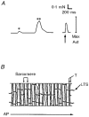
Figure 3. Spontaneous force responses and the longitudinal tubular system
A, spontaneous force responses (*13 % and **91 % of the maximum Ca2+-activated force (Max Act)) recorded in a skinned EDL fibre. Most of the length of the fibre (2.0 mm) must have been simultaneously activated in order to elicit near-maximal force, and even the small spontaneous response must have involved coordinated activation of many sarcomeres. The rise time of the larger spontaneous response (∼200 ms) shows that the contraction was synchronised by one or more action potentials travelling along the fibre at an effective longitudinal velocity of ∼10 mm s−1. Transverse field stimulation (arrow) evoked a twitch response with a rise time of ∼40 ms. Maximum force (Max Act) was determined as in Fig. 1. Segment diameter, 38 μm. B, schematic diagram of the transverse tubular system (T) and the associated longitudinal tubular system (LTS), based on electron micrographs of fast-twitch mammalian fibres in Franzini-Armstrong et al. (1988). The LTS evidently provides a pathway for action potentials (AP) to travel lengthways inside a fibre, ensuring synchronous activation of adjacent sarcomeres even if the action potential on the sarcolemma fails to penetrate the T-system.

Comment in

  • Chronicle of skinned muscle fibres.
    Stienen GJ. Stienen GJ. J Physiol. 2000 Aug 15;527 Pt 1(Pt 1):1. doi: 10.1111/j.1469-7793.2000.t01-2-00001.x. J Physiol. 2000. PMID: 10944165 Free PMC article. No abstract available.

References

    1. Allen DA, Lännergren J, Westerblad H. Muscle cell function during prolonged activity: cellular mechanisms of fatigue. Experimental Physiology. 1995;80:497–527. - PubMed
    1. Bezanilla F, Caputo C, Gonzalez-Serratos H, Venosa RA. Sodium dependence of the inward spread of activation in isolated twitch muscle fibres of the frog. The Journal of Physiology. 1972;223:507–523. - PMC - PubMed
    1. Chandler WK, Rakowski RF, Schneider MF. Effects of glycerol treatment and maintained depolarization on charge movement in skeletal muscle. The Journal of Physiology. 1976;254:285–316. - PMC - PubMed
    1. Costantin LL. The role of sodium current in the radial spread of contraction in frog muscle fibres. Journal of General Physiology. 1970;55:703–715. - PMC - PubMed
    1. Costantin LL, Podolsky RJ. Depolarization of the internal membrane system in the activation of frog skeletal muscle. Journal of General Physiology. 1967;50:1101–1124. - PMC - PubMed

Publication types

MeSH terms

LinkOut - more resources