. 2000 Aug 29;97(18):9925-30.
doi: 10.1073/pnas.160255397.

Directed evolution of a (beta alpha)8-barrel enzyme to catalyze related reactions in two different metabolic pathways

Affiliations

Directed evolution of a (beta alpha)8-barrel enzyme to catalyze related reactions in two different metabolic pathways

C Jürgens et al. Proc Natl Acad Sci U S A. .

Abstract

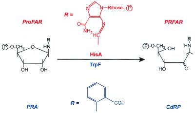
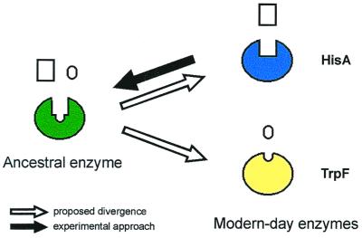
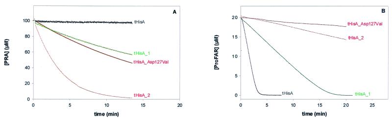
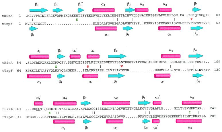
Enzymes participating in different metabolic pathways often have similar catalytic mechanisms and structures, suggesting their evolution from a common ancestral precursor enzyme. We sought to create a precursor-like enzyme for N'-[(5'-phosphoribosyl)formimino]-5-aminoimidazole-4-carboxamide ribonucleotide (ProFAR) isomerase (HisA; EC ) and phosphoribosylanthranilate (PRA) isomerase (TrpF; EC ), which catalyze similar reactions in the biosynthesis of the amino acids histidine and tryptophan and have a similar (betaalpha)(8)-barrel structure. Using random mutagenesis and selection, we generated several HisA variants that catalyze the TrpF reaction both in vivo and in vitro, and one of these variants retained significant HisA activity. A more detailed analysis revealed that a single amino acid exchange could establish TrpF activity on the HisA scaffold. These findings suggest that HisA and TrpF may have evolved from an ancestral enzyme of broader substrate specificity and underscore that (betaalpha)(8)-barrel enzymes are very suitable for the design of new catalytic activities.


Figures

Figure 1
Figure 1
HisA and TrpF catalyze similar reactions in histidine and tryptophan biosynthesis. HisA and TrpF catalyze the isomerizations of the aminoaldoses N′-[(5′-phosphoribosyl)formimino]-5-aminoimidazole-4-carboxamide ribonucleotide (ProFAR) and phosphoribosylanthranilate (PRA) to the aminoketoses N′-[(5′-phosphoribulosyl)formimino]-5-aminoimidazole-4-carboxamide ribonucleotide (PRFAR) and 1-(o-carboxyphenylamino)-1-deoxyribulose 5-phosphate (CdRP).
Figure 2
Figure 2
Experimental approach for testing the patchwork hypothesis (4) of enzyme evolution. Modern-day enzymes such as HisA and TrpF are highly specific catalysts that may have evolved from a common ancestor enzyme that was less specific. Starting from HisA, we tried to reverse the postulated evolutionary path, creating an enzyme capable of catalyzing both the HisA and the TrpF reaction.
Figure 3
Figure 3
TrpF (A) and HisA (B) activities of tHisA variants. (A) The TrpF reaction was monitored essentially as described (18). Each variant (21 μM) was incubated with 100 μM in situ synthesized PRA in the presence of 0.7 μM indoleglycerol-phosphate synthase from E. coli. The buffer conditions were 50 mM Tris⋅HCl, pH 7.5 at 25°C, containing 4 mM MgEDTA and 2 mM DTT. (B) The HisA reaction was monitored essentially as described (19). Each variant, 0.25 μM tHisA, 0.5 μM tHisA_1, 21 μM tHisA_2, or 21 μM tHisA_Asp127Val, was incubated with 20 μM enzymatically synthesized ProFAR (22) in the presence of 1 μM ImGP synthase from T. maritima and of 5 mM glutamine. The buffer conditions were 50 mM Tris⋅HCl, pH 7.5 at 25°C, containing 2 mM EDTA and 1 mM DTT.
Figure 4
Figure 4
Structure-based amino acid sequence alignment of tHisA and tTrpF. The backbone atoms from the x-ray structures of tHisA (8) and tTrpF (23) were superimposed with an rms deviation of 2.2 Å (24). β-Strands are represented by blue arrows, α-helices by red cylinders. Identical residues (10%) between tHisA and tTrpF are indicated by (|). Catalytically important residues of wild-type tHisA (Asp-8, Asp-127, and Thr-164; S. Schmidt, M. Henn-Sax, and R.S., unpublished data) and of wild-type tTrpF (Cys-7 and Asp-126; ref. 25) are in boldface. Residues involved in binding of the single phosphate moiety of PRA and of the two phosphate moieties of ProFAR are underlined. For tHisA, these residues were identified by a structure-based sequence alignment with the related tHisF protein, whose x-ray structure showed two phosphate ions bound to the active site (8). The acquired amino acids of tHisA_1 (green) and tHisA_2 (red) are given between the tHisA and the tTrpF sequences.

References

    1. Gerlt J A, Babbitt P C. Curr Opin Chem Biol. 1998;2:607–612. - PubMed
    1. Horowitz N H. Proc Natl Acad Sci USA. 1945;31:153–157. - PMC - PubMed
    1. Altamirano M M, Blackburn J M, Aguayo C, Fersht A R. Nature (London) 2000;403:617–622. - PubMed
    1. Jensen R A. Annu Rev Microbiol. 1976;30:409–425. - PubMed
    1. Winkler M E. In: Escherichia coli and Salmonella: Cellular and Molecular Biology. 2nd Ed. Neidhardt F C, Ingraham J L, Low K B, Magasanik B, Schaechter M, Umbarger H E, editors. Vol. 1. Washington, DC: Am. Soc. Microbiol.; 1996. pp. 485–505.

Publication types

Substances