. 2000 Aug 29;97(18):10002-7.
doi: 10.1073/pnas.170093297.

The human papillomavirus type 16 E6 and E7 oncoproteins cooperate to induce mitotic defects and genomic instability by uncoupling centrosome duplication from the cell division cycle

Affiliations

The human papillomavirus type 16 E6 and E7 oncoproteins cooperate to induce mitotic defects and genomic instability by uncoupling centrosome duplication from the cell division cycle

S Duensing et al. Proc Natl Acad Sci U S A. .

Abstract

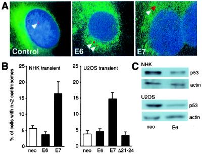
Loss of genomic integrity is a defining feature of many human malignancies, including human papillomavirus (HPV)-associated preinvasive and invasive genital squamous lesions. Here we show that aberrant mitotic spindle pole formation caused by abnormal centrosome numbers represents an important mechanism in accounting for numeric chromosomal alterations in HPV-associated carcinogenesis. Similar to what we found in histopathological specimens, HPV-16 E6 and E7 oncoproteins cooperate to induce abnormal centrosome numbers, aberrant mitotic spindle pole formation, and genomic instability. The low-risk HPV-6 E6 and E7 proteins did not induce such abnormalities. Whereas the HPV-16 E6 oncoprotein has no immediate effects on centrosome numbers, HPV-16 E7 rapidly induces abnormal centrosome duplication. Thus our results suggest a model whereby HPV-16 E7 induces centrosome-related mitotic disturbances that are potentiated by HPV-16 E6.


Figures

Figure 1
Figure 1
(A) Mitotic figures with abnormal numbers of centrosomes in both preinvasive (squamous intraepithelial lesions of the cervix, SIL) and invasive (vulvar squamous cell carcinoma, SCC) HPV-associated genital squamous lesions. Centrosomes were visualized by immunofluorescence staining for pericentrin. Nuclei were visualized with Hoechst 33258 DNA dye. Pictures were merged to show the arrangement of centrosomes and mitotic figures. Each of the examples shows a tripolar arrangement of spindle poles. In addition, focusing up and down revealed individual spindle poles consisting of more than one centrosome (arrowheads). (B) (Left) Immunoblot analysis revealed decreased levels of p53 in HPV-16 E6-expressing NHKs. (Right) Immunoblot detection of HPV-16 E7 protein in NHKs with stable expression of this protein. (C) Centrosome abnormalities in HPV-16 E6-, E7-, and E6/E7-expressing NHKs. Centrosomes were visualized by immunofluorescence staining for γ-tubulin (arrowheads), and nuclei were visualized with Hoechst 33258 DNA dye. LXSN-vector-infected keratinocytes (NHK) are shown as the control. Pictures were acquired with a multiband filter set. (D) Quantitation of centrosomal abnormalities in high-risk HPV-16 E6-, E7-, E6/E7-, and low-risk HPV-6 E6 or E7-expressing NHK clones. LXSN-vector infected keratinocytes are shown as the control. Bar graphs show the mean ± SEM of at least triplicate centrosome quantitations.
Figure 2
Figure 2
(A) NHKs were transiently transfected with HPV-16 E6 or E7 and a vector encoding farnesylatable GFP. Only GFP-positive cells were evaluated. Keratinocytes transfected with the parental plasmid were used as the control. Centrosomes were visualized by immunofluorescence staining for γ-tubulin (arrowheads). Nuclei were counterstained with the DNA dye Hoechst 33258. Pictures were obtained with a multiband filter set. (B) Quantitation of centrosomal abnormalities in NHKs and U2OS cells transiently transfected with HPV-16 E6 or E7. Cells transfected with the parental plasmid (neo) were used as the control. Bar graphs show the mean ± SEM of three independent experiments. (C) Decreased p53 levels in response to transient HPV-16 E6 expression in NHKs (Upper) and U2OS cells (Lower). An actin blot is shown to demonstrate equal loading.
Figure 3
Figure 3
(A) Immunoblot detection of E7 in HPV-16 E7-expressing U2OS cells. (B) Quantitation of centrosomal abnormalities in HPV-16 E7-expressing U2OS cells. Empty vector (neo) transfected cells are shown as the control. Bar graphs show the mean ± SEM of three independent experiments. (C) Ultrastructural analysis of centrosomes in HPV-16 E7-expressing U2OS cells, using transmission electron microscopy. Increased procentriole formation (arrowhead) was observed in E7-expressing U2OS cells compared with matched controls. See text for details. (D) (Left) Treatment of E7-expressing U2OS cells with the cdk2 inhibitor roscovitine decreases the proportion of cells with abnormal centrosome numbers within 24 h. Controls denote E7-expressing U2OS cells treated with solvent (DMSO). (Center) Coexpression of HPV-16 E7 and dominant-negative mutant cdk2 (dn-cdk2) in U2OS cells. (Right) Coexpression of HPV-16 E7 and dominant-negative mutant DP1 (dn-DP1) in U2OS cells. Cell populations transfected with the parental plasmid (neo) are shown as controls. Bar graphs show the mean ± SEM of four independent experiments.
Figure 4
Figure 4
(A) Abnormal metaphases with multiple participating centrosomes in HPV-16 E6-, E7-, or E6/E7-expressing NHKs. LXSN vector-infected cells are shown as the control. Centrosomes were visualized by immunofluorescence staining for γ-tubulin, and nuclei were visualized with the DNA dye Hoechst 33258. (B) Quantitation of metaphase cells deviant from a bipolar centrosome arrangement in HPV-16 E6-, E7-, or E6/E7-expressing stable clones of NHKs (Left) and U2OS cells transiently transfected with HPV-16 E6, E7, or mutant HPV-16 E7Δ21–24 (Right). LXSN-vector-infected keratinocytes and U2OS cells transfected with the parental plasmid (neo) are shown as controls. The bar graph denotes the result of one representative experiment; at least 100 metaphases were analyzed for each sample. (C) Control anaphase and anaphase of a HPV-16 E6/E7-expressing NHK cell displaying asymmetrical distribution of the condensed chromosomes, a tripolar centrosome arrangement, and one spindle pole with two centrosomes (arrowhead).
Figure 5
Figure 5
(A) Fluorescence in situ hybridization analysis of chromosome 11 in HPV-16 E7-expressing NHKs. Nuclei were counterstained with propidium iodide. Control cells represent matched cultures infected with the parental LXSN vector. (B) Quantitation of chromosome 11 copy numbers in NHKs stably expressing HPV-16 E6, E7, or both viral oncoproteins. LXSN vector-infected keratinocytes are shown as controls.

References

    1. zur Hausen H. Science. 1991;254:1167–1173. - PubMed
    1. Solinas-Toldo S, Dürst M, Lichter P. Proc Natl Acad Sci USA. 1997;94:3854–3859. - PMC - PubMed
    1. Heselmeyer K, Schröck E, Du Manoir S, Blegen H, Shah K, Steinbeck R, Auer G, Ried T. Proc Natl Acad Sci USA. 1996;93:479–484. - PMC - PubMed
    1. Hashida T, Yasumoto S. J Gen Virol. 1991;72:1569–1577. - PubMed
    1. White A E, Livanos E M, Tlsty T D. Genes Dev. 1994;8:666–677. - PubMed

Publication types

MeSH terms

Substances

LinkOut - more resources