. 2000 Aug 29;97(18):10107-12.
doi: 10.1073/pnas.170295097.

TOM1, an Arabidopsis gene required for efficient multiplication of a tobamovirus, encodes a putative transmembrane protein

Affiliations

TOM1, an Arabidopsis gene required for efficient multiplication of a tobamovirus, encodes a putative transmembrane protein

T Yamanaka et al. Proc Natl Acad Sci U S A. .

Abstract

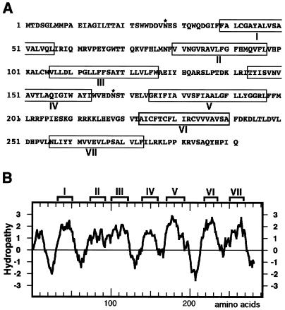
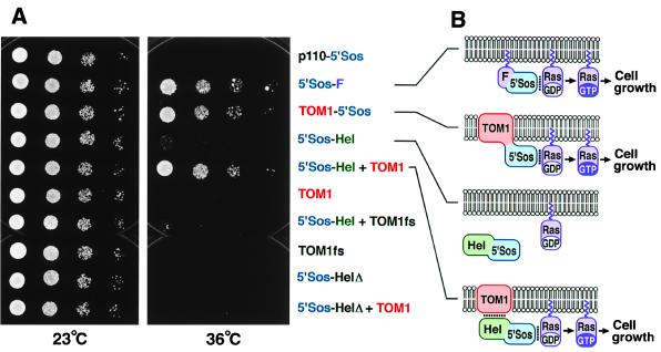
Host-encoded factors play an important role in virus multiplication, acting in concert with virus-encoded factors. However, information regarding the host factors involved in this process is limited. Here we report the map-based cloning of an Arabidopsis thaliana gene, TOM1, which is necessary for the efficient multiplication of tobamoviruses, positive-strand RNA viruses infecting a wide variety of plants. The TOM1 mRNA is suggested to encode a 291-aa polypeptide that is predicted to be a multipass transmembrane protein. The Sos recruitment assay supported the hypothesis that TOM1 is associated with membranes, and in addition, that TOM1 interacts with the helicase domain of tobamovirus-encoded replication proteins. Taken into account that the tobamovirus replication complex is associated with membranes, we propose that TOM1 participates in the in vivo formation of the replication complex by serving as a membrane anchor.


Figures

Figure 1
Figure 1
Genetic mapping and positional cloning of TOM1. (A) Genetic map around the TOM1 locus on chromosome 4. Vertical bars represent DNA markers. Each number represents recombination events between the marker and the TOM1 locus among 3,103 F2 plants. (B and C) Contigs of ESSA II bacterial artificial chromosome clones and Mitsui P1 clones encompassing the TOM1 locus, respectively. (D) Fine map around the TOM1 locus. Vertical bars and numbers are the same as in A. The position and orientation of the TOM1 gene is shown by the arrow. (E) T-DNA contigs. T-DNA clones derived from the P1 clones 8C3 or 5F6 were organized into an overlapping set that spanned the TOM1 locus. These T-DNA clones were used to genetically transform tom1–1 mutant plants, which subsequently were tested for complementation of the TMV-Cg multiplication phenotype. The ratio of numbers of transformed lines that showed wild-type level TMV-Cg multiplication vs. total lines tested for TMV-Cg multiplication is indicated at the right of each T-DNA clone. (F) Intron-exon organization of the TOM1 gene and the tom1 mutations. Exons are indicated by boxes. Open boxes indicate noncoding regions, and filled boxes indicate coding regions. Mutations in the three tom1 alleles are shown below the intron/exon structure.
Figure 2
Figure 2
Complementation of tom1 mutation with T-DNA clone pYT1. (A) Cosegregation of T-DNA and complementation. T2 progenies derived from a tom1–1 mutant transformed with T-DNA clone pYT1 were inoculated with TMV-Cg. Two weeks after inoculation, total protein was prepared from the inoculated plants, separated by SDS/PAGE, and stained with Coomassie brilliant blue (Upper). For each T2 plant, the NPTII sequence was amplified from purified genomic DNA by PCR with specific primer sets and analyzed by agarose gel electrophoresis followed by ethidium bromide staining (Lower). The positions of TMV-Cg CP and the PCR-amplified NPTII DNA fragment are indicated. (B) Complementation at protoplast level. Direct descendants of a T2 progeny derived from a tom1–1 mutant transformed with T-DNA clone pYT1 and carrying the transgene homozygously were used to prepare protoplasts. Protoplasts were inoculated with TMV-Cg RNA by electroporation, cultured for 4, 10, or 20 h, and the accumulation of TMV-Cg-related RNAs was examined by Northern blot hybridization. The position of genomic RNA (G), subgenomic mRNAs for 30-kDa protein (30K), and CP are indicated at the right. As controls, analysis with nontransformed Col-0 (wt) and tom1–1 (tom1) protoplasts were performed in parallel.
Figure 3
Figure 3
Northern blot hybridization analysis of the TOM1 mRNA. (A) TOM1 mRNA accumulation in wild-type and mutant plants. Total RNA was extracted from aerial tissues of 25-day-old noninoculated wild-type (Col-0 and WS) and mutant (tom1–1, tom1–2, and tom1–3) plants. Note that tom1–1 and tom1–2 are derived from Col-0, whereas tom1–3 is from WS (see Materials and Methods). RNA samples (10 μg) were denatured by glyoxal, separated by 1% agarose gel electrophoresis, and blotted onto a nylon membrane. Duplicate blots were prepared and probed with 32P-labeled DNAs hybridizing with either TOM1 or 18S rRNA (43) sequences. To prepare the TOM1-specific probe, a DNA fragment corresponding to the predicted TOM1 ORF was amplified by PCR from a cDNA clone and gel purified. (B) TOM1 mRNA levels are not altered by TMV-Cg infection. Total RNA was extracted from aerial tissues of mock-inoculated (mock) and TMV-Cg-inoculated (TMV) Col-0 plants. Twenty-day-old plants were inoculated and samples were harvested 3 or 7 days after inoculation. Northern blot hybridization was performed as in A. The positions of TOM1 mRNA and 18S rRNA are indicated (A Left). The positions of CMV (Y-strain) RNA3 (2,215 nt) and RNA4 (1,033 nt) used as size markers are shown (B Right).
Figure 4
Figure 4
Structure of TOM1. (A) Deduced amino acid sequence of TOM1. Boxed amino acid residues represent those belonging to putative membrane-spanning regions, as predicted by the sosui program (29). The program phdthtm (30) did not list the region VII, and psort (31) did not list the region II as a transmembrane region. Asn residues marked by * are putative glycosylation sites. (B) Hydropathy plot for the deduced amino acid sequence of TOM1. The hydropathy plot was created by the method described by Kyte and Doolittle (44). The regions predicted to be membrane spanning in A are indicated above the plot.
Figure 5
Figure 5
Interaction between TOM1 and the TMV-Cg-encoded replication proteins. (A) cdc25–2 yeast strains harboring plasmids designed to constitutively express indicated proteins were diluted in sterile water to absorbance at 600 nm of 0.2, 0.025, 0.003, and 0.0004 (8-fold serial dilutions). Each dilution (2 μl) was spotted onto YAPD plates [1% (wt/vol) yeast extract/2% (wt/vol) peptone/2% (wt/vol) glucose/0.004% (wt/vol) adenine hemisulfate/2% (wt/vol) agar] and cultured at 23°C for 52 h or 36°C for 66 h. Part of p110 β, a subunit of phosphatidylinositol-3-phosphate kinase, fused to 5′Sos (p110–5′Sos), and 5′Sos fused to Ras farnesylation signal (5′Sos-F) are negative and positive controls, respectively, for suppression of cdc25–2 temperature sensitivity (24). TOM1fs and 5′Sos-HelΔ are frameshifting and deletion derivatives of TOM1 and 5′Sos-Hel, respectively. (B) Models explaining the results of A. Lipid bilayers indicate plasma membranes with the lower sides cytoplasmic. Noncovalent interactions are indicated by dotted lines. Covalent linkage of 5′Sos polypeptide with TOM1 or a noncovalent interaction between TOM1 and the helicase domain of TMV-Cg-encoded replication proteins in 5′Sos-Hel recruits 5′Sos to the plasma membrane to activate Ras signaling.

References

    1. Buck K W. Adv Virus Res. 1996;47:159–251. - PMC - PubMed
    1. Lai M M C. Virology. 1998;244:1–12. - PubMed
    1. Quadt R, Kao C C, Browning K S, Hershberger R P, Ahlquist P. Proc Natl Acad Sci USA. 1993;90:1498–1502. - PMC - PubMed
    1. Osman T A, Buck K W. J Virol. 1997;71:6075–6082. - PMC - PubMed
    1. Pardigon N, Strauss J H. J Virol. 1996;70:1173–1181. - PMC - PubMed

Publication types

Associated data