. 2000 Aug 15;97(17):9443-8.
doi: 10.1073/pnas.97.17.9443.

Intramolecular control of transcriptional activity by the NK2-specific domain in NK-2 homeodomain proteins

Affiliations

Intramolecular control of transcriptional activity by the NK2-specific domain in NK-2 homeodomain proteins

H Watada et al. Proc Natl Acad Sci U S A. .

Abstract

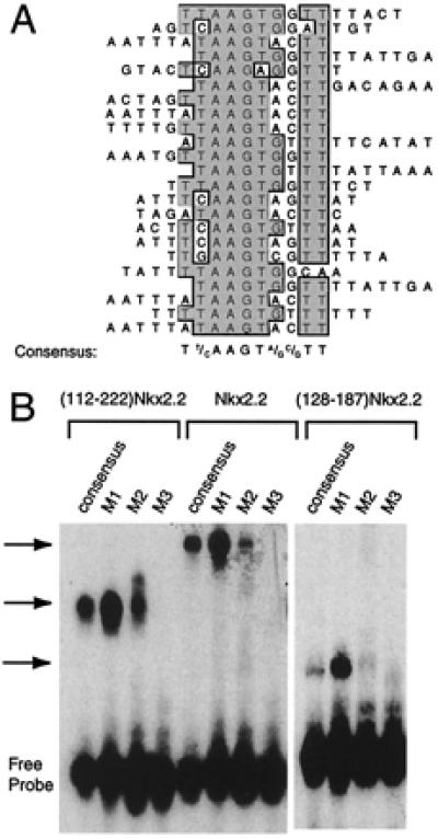
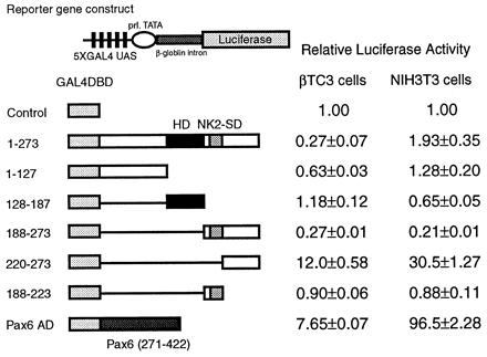
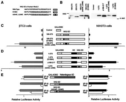
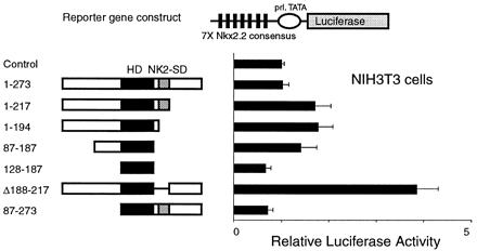
The developmentally important homeodomain transcription factors of the NK-2 class contain a highly conserved region, the NK2-specific domain (NK2-SD). The function of this domain, however, remains unknown. The primary structure of the NK2-SD suggests that it might function as an accessory DNA-binding domain or as a protein-protein interaction interface. To assess the possibility that the NK2-SD may contribute to DNA-binding specificity, we used a PCR-based approach to identify a consensus DNA-binding sequences for Nkx2.2, an NK-2 family member involved in pancreas and central nervous system development. The consensus sequence (T(C)(T)AAGT(G)(A)(G)(C)TT) is similar to the known binding sequences for other NK-2 homeodomain proteins, but we show that the NK2-SD does not contribute significantly to specific DNA binding to this sequence. To determine whether the NK2-SD contributes to transactivation, we used GAL4-Nkx2. 2 fusion constructs to map a powerful transcriptional activation domain in the C-terminal region beyond the conserved NK2-SD. Interestingly, this C-terminal region functions as a transcriptional activator only in the absence of an intact NK2-SD. The NK2-SD also can mask transactivation from the paired homeodomain transcription factor Pax6, but it has no effect on transcription by itself. These results demonstrate that the NK2-SD functions as an intramolecular regulator of the C-terminal activation domain in Nkx2.2 and support a model in which interactions through the NK2-SD regulate the ability of NK-2-class proteins to activate specific genes during development.


Figures

Figure 1
Figure 1
Nkx2.2 binding site selection. (A) Binding site selection was performed with the human Nkx2.2 homeodomain and NK2-SD (amino acids 112–221) and a set of oligonucleotides containing a 15-bp random stretch flanked by PCR primer sites. Sequenced products from the eighth round of selection are shown aligned by the clustal w alignment algorithm in macvector 6.5 software (Oxford Molecular). The consensus sequence that emerges from the best-fit line-up is shown. (B) An EMSA using in vitro translated ()Nkx2.2, full-length Nkx2.2, and the Nkx2.2 homeodomain alone (amino acids 128–187). Different 32P-labeled oligonucleotides (sequences shown in Table 1) were incubated with 1 μl of each in vitro translated protein for 15 min at room temperature and then subjected to electrophoresis on a 5% polyacrylamide gel. The free probe and retarded complex are indicated.
Figure 2
Figure 2
The transactivation domain of Nkx2.2 maps to the C terminus. A reporter plasmid containing five tandem copies of the GAL4 UAS upstream of the E1b minimal promoter driving luciferase and an expression plasmid encoding each GAL4DBD-Nkx2.2 fusion construct were cotransfected with a CMV promoter-driven β-galactosidase expression plasmid. All luciferase activities are corrected for β-galactosidase activity. Relative luciferase activities are calculated, with the activity of cells transfected with the GAL4DBD alone set at 1. All data are shown as mean ± SEM. (A) The reporter plasmid is indicated schematically. (B) The results of the one-hybrid assays. (Left) Results from βTC3 cells. (Right) Results from NIH 3T3 cells. (C) Results of detailed mapping of the activation domain. (Left) Results from βTC3 cells. (Right) Results from NIH 3T3 cells.
Figure 3
Figure 3
Full-length Nkx2.2 acts as a weak repressor in βTC3 cells. A reporter plasmid containing five tandem copies of the GAL4 UAS upstream of the prolactin minimal promoter driving luciferase and an expression plasmid encoding each GAL4DBD-Nkx2.2 fusion construct or a GAL4DBD-Pax6 activation domain fusion construct were cotransfected with CMVβgal. All luciferase activities are corrected for β-galactosidase activity. Relative luciferase activities are calculated, with the activity of cells transfected with the GAL4DBD alone set at 1. All data are shown as mean ± SEM.
Figure 4
Figure 4
Characterization of the transactivation domain of Nkx2.2. A reporter plasmid containing five tandem copies of the GAL4 UAS upstream of the E1b minimal promoter driving luciferase and an expression plasmid encoding each GAL4DBD-Nkx2.2 fusion construct were cotransfected with a CMV promoter-driven β-galactosidase expression plasmid. All luciferase activities are corrected for β-galactosidase activity. All data are shown as mean ± SEM. (A) The sequences of the native and mutant NK2-SD. The NK2-SD is underlined, and mutations are in boldface. (B) Western blotting data. Each construct is transfected into NIH 3T3 cells. Five micrograms of nuclear extracts from each cell is loaded. (C) The relative luciferase activities from βTC3 cells and NIH 3T3 cells transfected with expression plasmids encoding the C-terminal region with wild-type or mutant NK2-SD fused to the GAL4DBD. Relative luciferase activities are calculated, with the activity of cells transfected with the GAL4DBD alone set at 1. (D) The relative luciferase activities from βTC3 cells and NIH 3T3 cells transfected with expression plasmids encoding the full-length Nkx2.2 with wild-type or mutant NK2-SD fused to the GAL4DBD. Relative luciferase activities are calculated, with the activity of cells transfected with GAL4DBD–full-length Nkx2.2 alone set at 1. Comparison with the GAL4DBD alone is shown in Fig. 2B. (E) Relative luciferase activities from cells transfected with the VP16 activation domain or Pax6 activation domain fused to wild-type or mutant NK2-SD and the GAL4DBD. Relative luciferase activities are calculated, with the activity of cells transfected with an expression plasmid containing the isolated VP16 activation domain fused to the GAL4DBD set at 1. VP16 activation domain fused to the GAL4DBD showed 516,000-fold activation in NIH 3T3 cells and 5,990-fold activation in βTC3 cells compared with the GAL4DBD alone. On the other hand, Pax6 activation domain fused to the GAL4DBD showed 102-fold activation in NIH 3T3 cells and 6.1-fold activation in βTC3 cells compared with the GAL4DBD alone.
Figure 5
Figure 5
Removal of the NK2-SD activates Nkx2.2. A reporter plasmid containing seven tandem copies of the Nkx2.2 DNA-binding consensus site (shown in Table 1) upstream of the prolactin promoter driving luciferase (pFOxLuc1–7XNk2) was cotransfected with the CMV promoter-driven expression plasmids encoding the Nkx2.2 cDNA shown. Relative luciferase activities are calculated, with the activity of cells transfected with a CMV expression plasmid without cDNA insert set at 1. All data are shown as mean ± SEM.

References

    1. Harvey R P. Dev Biol. 1996;178:203–216. - PubMed
    1. Kimura S, Hara Y, Pineau T, Fernandez-Salguero P, Fox C H, Ward J M, Gonzalez F J. Genes Dev. 1996;10:60–69. - PubMed
    1. Sussel L, Marin O, Kimura S, Rubenstein J L. Development (Cambridge, UK) 1999;126:3359–3370. - PubMed
    1. Lints T J, Parsons L M, Hartley L, Lyons I, Harvey R P. Development (Cambridge, UK) 1993;119:419–431. - PubMed
    1. Biben C, Harvey R P. Genes Dev. 1997;11:1357–1369. - PubMed

MeSH terms

LinkOut - more resources