. 2000 Aug 15;97(17):9579-84.
doi: 10.1073/pnas.97.17.9579.

Multiple functional defects in peripheral autonomic organs in mice lacking muscarinic acetylcholine receptor gene for the M3 subtype

Affiliations

Multiple functional defects in peripheral autonomic organs in mice lacking muscarinic acetylcholine receptor gene for the M3 subtype

M Matsui et al. Proc Natl Acad Sci U S A. .

Abstract

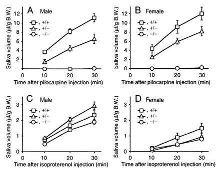
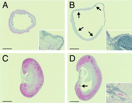
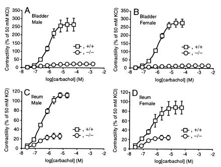
Muscarinic acetylcholine receptors consist of five distinct subtypes and have been important targets for drug development. In the periphery, muscarinic acetylcholine receptors mediate cholinergic signals to autonomic organs, but specific physiological functions of each subtype remain poorly elucidated. Here, we have constructed and analyzed mutant mice lacking the M(3) receptor and have demonstrated that this subtype plays key roles in salivary secretion, pupillary constriction, and bladder detrusor contractions. However, M(3)-mediated signals in digestive and reproductive organs are dispensable, likely because of redundant mechanisms through other muscarinic acetylcholine receptor subtypes or other mediators. In addition, we have found prominent urinary retention only in the male, which indicates a considerable sex difference in the micturition mechanism. Accordingly, this mutant mouse should provide a useful animal model for investigation of human diseases that are affected in the peripheral cholinergic functions.


Figures

Figure 1
Figure 1
Generation of Chrm3-deficient mice. (A) Targeting strategy by homologous recombination in ES cells. Targeting vector pChrm3-N2 contained the neo gene and the diphtheria toxin α-subunit gene (DTA; ref. 10) driven by the phosphoglycerate kinase I promoter. Arrows MF11 and PR2 indicate the PCR primers used for homologous recombinant screening, whereas MF28, AF2, and MR24 for genotyping. BamHI (B), HindIII (H), and EcoRI (R) sites relevant to the identification of homologous recombinant ES cell clones are shown together with the expected sizes hybridizable to the Chrm3 and the neo probes. (B and C) Confirmation of the homologous recombination in ES cell clones by Southern hybridization. (B) Hybridization with the Chrm3 probe showing a 4.2-kb band specific to the targeted allele and a 7.0-kb band derived from the wild-type allele. (C) Hybridization with the neo probe showing a 5.3-kb band specific to the targeted allele. (D) Northern analysis showing mRNA levels of Chrm1 and Chrm3 in the brains of wild-type, heterozygous, and homozygous mice. Note that the Chrm3 mRNA in the heterozygous brain decreased to about half of the wild-type brain and is absent in the homozygous brain. The mRNA levels of Chrm1 were not different among the three genotypes. (Bottom) Signals hybridized with a Gapd probe used as an internal control.
Figure 2
Figure 2
Growth curves of male (A) and female (B) mice. The value of each point is the average body weight (±SEM) of the wild-type and homozygous mutant mice (n = 5 to 7) monitored for 15 weeks. When fed a standard pellet diet, the growth of the homozygous mice (○) was retarded significantly, compared with that of the wild-type mice (□). Asterisks (*) indicate body weights 70% or less than those of wild-type mice. In both sexes, the weight differences became noticeable around 3 weeks of age and were most prominent at 4–5 weeks. When fed with hydrated paste diet from 2 weeks of age, the growth of the homozygous pups (●) improved significantly.
Figure 3
Figure 3
Appearance of pupils of the heterozygous (A and C) and homozygous (B and D) mutant mice (4-month old female littermates) on instillation of an antagonist (atropine) under bright illumination. (A and B) Before instillation. The pupils of the homozygotes (B) are larger than those of the heterozygous mouse (A). (C and D) Thirty minutes after an instillation of 1% atropine. Instillation of atropine caused full dilatation of the pupils not only in the heterozygous (C) but also in the homozygous mouse (D). Bars = 1 mm.
Figure 4
Figure 4
Stimulated salivary responses. (A) Response in the male mice on stimulation with pilocarpine. (B) Response in female mice on stimulation with pilocarpine. (C) Response in male mice on stimulation with isoproterenol. (D) Response in the female mice on stimulation with isoproterenol. Time courses of cumulative salivary volume (mean ± SEM) were shown that were determined at 10, 20, and 30 min after each stimulation. □, ▵, and ○ indicate the data of wild-type, heterozygous, and homozygous mice, respectively. Each symbol represents the data of four to five experiments.
Figure 5
Figure 5
Histology of the bladder and kidney. (A and B) Male urinary bladders of wild-type (A) and homozygous (B) mice. The bladder of the homozygous mouse is distended with focal submucosal infiltrations of lymphocytes (arrows). Note that the smooth muscle layer is thinner, especially in the region of right upper quadrant, because of severe distension. Inset (×50 higher magnification) shows the normal epithelium with a submucosal lymphocyte infiltration. (C and D) Kidneys of wild-type (C) and homozygous (D) male mice. The renal calix of the homozygous mice is distended. Inset (×10) shows a representative lymphocyte infiltration in the perivascular region in the medulla (arrow). Bars = 1.5 mm (A and B) and 2.0 mm (C and D).
Figure 6
Figure 6
Reduced bladder (A and B) and ileal (C and D) smooth muscle contractions in response to carbachol in the homozygous mice. Responses to carbachol are shown (mean ± SEM) as percentages of those to 50 mM KCl in male (A and C) wild-type (n = 4) and homozygous (n = 4) mice and female (B and D) wild-type (n = 4) and homozygous (n = 4) mice. □ and ○ indicate the data of wild-type and homozygous mice, respectively.

References

    1. Mitchelson F. Pharmacol Ther. 1988;37:357–423. - PubMed
    1. Caulfield M P, Birdsall N J M. Pharmacol Rev. 1998;50:279–290. - PubMed
    1. Matsui M, Araki Y, Karasawa H, Matsubara N, Taketo M M, Seldin M F. Genes Genet Syst. 1999;74:15–21. - PubMed
    1. Ehlert F J, Sawyer G W, Esqueda E E. Life Sci. 1999;64:387–394. - PubMed
    1. Eglen R M, Choppin A, Dillon M P, Hegde S. Curr Opin Chem Biol. 1999;3:426–432. - PubMed

Publication types

MeSH terms