. 2000 Sep;68(9):5277-83.
doi: 10.1128/IAI.68.9.5277-5283.2000.

ankA: an Ehrlichia phagocytophila group gene encoding a cytoplasmic protein antigen with ankyrin repeats

Affiliations

ankA: an Ehrlichia phagocytophila group gene encoding a cytoplasmic protein antigen with ankyrin repeats

P Caturegli et al. Infect Immun. 2000 Sep.

Abstract

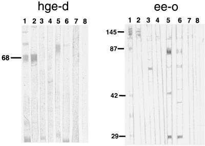
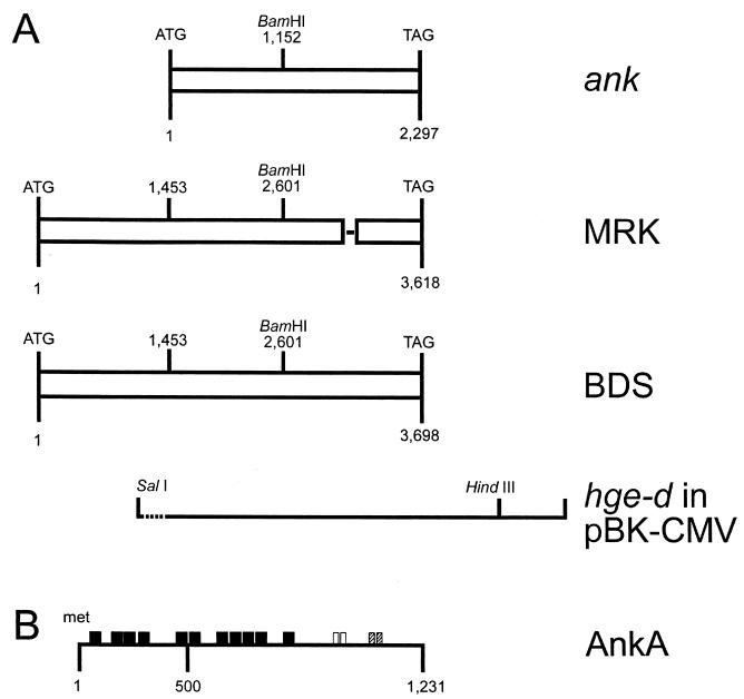
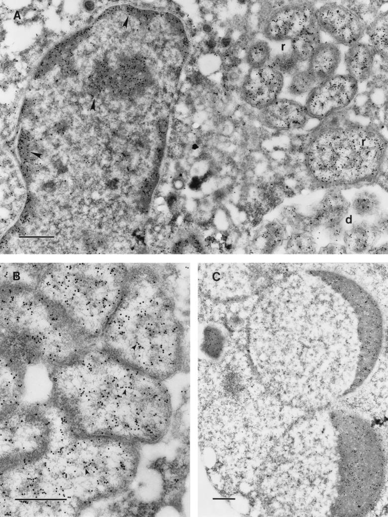
Human granulocytic ehrlichiosis (HGE) is a potentially fatal, tick-borne disease caused by a bacterium related or identical to Ehrlichia phagocytophila. To identify and characterize E. phagocytophila group-specific protein antigen genes, we prepared and screened HGE agent and Ehrlichia equi genomic DNA expression libraries using polyclonal equine E. equi antibodies. Two clones, one each from HGE agent and E. equi, that were recognized specifically by antibodies to the E. phagocytophila group ehrlichiae had complete open reading frames of 3,693 and 3,615 nucleotides, respectively. The two clones were 96.6% identical and predicted a protein with at least 11 tandemly repeated ankyrin motifs. Thus, the gene was named ank (for ankyrin). When the encoded protein, named AnkA, was expressed in Escherichia coli, it was recognized by antibodies from rabbits and mice immunized with the HGE agent, sera from humans convalescent from HGE, and sera from horses convalescent from HGE and E. equi infection. Monospecific AnkA antibodies reacted with proteins in HGE agent immunoblots, and AnkA monoclonal antibodies detected cytoplasmic antigen in E. phagocytophila group bacteria and also detected antigen associated with chromatin in infected but not uninfected HL-60 cell cultures. These results suggest that this Ehrlichia protein may influence host cell gene expression.


Figures

FIG. 1
FIG. 1
E. coli transformed with hge-d (left panel) or ee-o (right panel) expresses a 68- or 145-kDa antigen, respectively, as determined by protein immunoblotting. The blots were reacted with the following primary antibodies: human anti-HGE agent (lanes 1), horse anti-HGE agent BDS strain (lanes 2), human anti-E. chaffeensis (lanes 3), dog anti-E. canis (lanes 4), human anti-Borrelia burgdorferi (lanes 5), and human anti-Babesia microti (lanes 6). Lanes 7 and 8 were reacted with normal human and horse sera, respectively. The location of the 68-kDa protein is indicated on the left.
FIG. 2
FIG. 2
(A) Schematic representation and alignment of HGE agent (strain BDS) and E. equi (strain MRK) AnkA open reading frames. For comparison, the previously reported ank gene (24) is also shown at the top. hge-d in pBK-CMV refers to the initial clone, highlighting the SalI and HindIII sites used to subclone into the pMAL-c2 expression vector; the dotted line represents 15 nt of vector sequence. The SalI-BamHI fragment was the one used for probing Southern blots. The single line within the MRK sequences identifies the approximate position of the 81 nt coding for the second 27-amino-acid repeat, which is missing in AnkA from E. equi. (B) Schematic representation of AnkA. Closed boxes, 11 ankyrin repeats; open boxes, two contiguous 27-amino-acid repeats; hatched boxes, two 11-amino-acid repeats. Gene, clone, and protein designations are shown on the right.
FIG. 3
FIG. 3
Southern blot analysis of restriction enzyme-digested DNA from the HGE agent, E. coli XLOLR, and HL-60 cells. Lane 1, λDNA BstEII marker; lane 2, 50 pg of hge-d linearized with BamHI; lanes 3 to 6, HGE agent genomic DNA digested with EcoRI (lane 3), HindIII (lane 4), XhoI (lane 5), and BglII (lane 6); lanes 7 to 9, PstI-digested genomic DNA from the HGE agent (lane 7), E. coli XLOLR (lane 8), and HL-60 cells (lane 9); lanes 10 to 12: BamHI-digested genomic DNA from the HGE agent (lane 10), E. coli XLOLR (lane 11), and HL-60 cells (lane 12); lanes 13 to 15: KpnI-digested genomic DNA from the HGE agent (lane 13), E. coli XLOLR (lane 14), and HL-60 cells (lane 15). Molecular sizes are shown on the left.
FIG. 4
FIG. 4
Protein immunoblots of recombinant MBP-AnkA (A), total lysate of E. coli transformed by ee-o (B), and whole HGE agent (C). (A) Recombinant MBP-AnkA probed with the following primary antibodies: mouse polyclonal anti-MBP-AnkA (lane 1), mouse polyclonal anti-HGE agent (BDS strain) (lane 2), nonimmune mouse serum (lane 3), rabbit anti-MBP (lane 4), nonimmune rabbit serum (lane 5), mouse monoclonal IE3 [IgG1(κ)] to MBP-AnkA (lane 6), and mouse monoclonal IgG1(κ) control antibody (lane 7). (B) Lysates of E. coli transformed by ee-o (lanes 1 and 3) or not transformed (lanes 2 and 4) were processed as described for panel A and probed with the following primary antibodies: mouse monoclonal IE3 [IgG1(κ)] to MBP-AnkA (lanes 1 and 2) and mouse polyclonal anti-HGE agent (lanes 3 and 4). (C) Whole HGE agent was processed as for panels A and B and probed with the following primary antibodies: rabbit anti-HGE agent (Webster strain) (lane 1), rabbit anti-MBP (lane 2), nonimmune rabbit serum (lane 3), mouse polyclonal anti-HGE agent (BDS strain) (lane 4), mouse polyclonal anti-MBP-AnkA (lane 5), and nonimmune mouse serum (lane 6). The numbers on the right represent the molecular masses (MWs) of HGE agent proteins that contain AnkA antigens (150, 90, 75, and 51 kDa) and the 42-kDa immunodominant HGE agent antigen that reacts only with polyclonal HGE agent antibodies.
FIG. 5
FIG. 5
Immunoelectron microscopic visualization of AnkA with monoclonal antibody IE3 in HL-60 cells infected with HGE agent (Webster strain). Bars, 0.5 μm. (A) The label is localized in the cytoplasm of the ehrlichiae, both reticulate (r) and dense-cored (d) cells, and on condensed chromatin of the host cell nucleus (arrowheads). (B) In reticulate cells, the cytoplasm is labeled and many gold particles are aligned along the DNA fibrils of the nucleoid (arrowheads). (C) In an apoptotic cell, condensed chromatin of the apoptotic nucleus is heavily labeled.

References

    1. Aguero-Rosenfeld M E, Horowitz H W, Wormser G P, McKenna D F, Nowakowski J, Munoz J, Dumler J S. Human granulocytic ehrlichiosis (HGE): a case series from a single medical center in New York State. Ann Intern Med. 1996;125:904–908. - PubMed
    1. Anderson B E, Dawson J E, Jones D C, Wilson K H. Ehrlichia chaffeensis, a new species associated with human ehrlichiosis. J Clin Microbiol. 1991;29:2838–2842. - PMC - PubMed
    1. Anderson B E, McDonald G A, Jones D C, Regnery R L. A protective protein antigen of Rickettsia rickettsii has tandemly repeated, near-identical sequences. Infect Immun. 1990;58:2760–2769. - PMC - PubMed
    1. Asanovich K M, Bakken J S, Madigan J E, Aguero-Rosenfeld M, Wormser G P, Dumler J S. Antigenic diversity of granulocytic Ehrlichia species isolates from humans in Wisconsin, New York, and a California horse. J Infect Dis. 1997;176:1029–1034. - PubMed
    1. Baeuerle P A, Baltimore D. NF-kappa B: ten years after. Cell. 1996;87:13–20. - PubMed

Publication types