. 2000 Sep;68(9):5377-84.
doi: 10.1128/IAI.68.9.5377-5384.2000.

Burkholderia pseudomallei induces cell fusion and actin-associated membrane protrusion: a possible mechanism for cell-to-cell spreading

Affiliations

Burkholderia pseudomallei induces cell fusion and actin-associated membrane protrusion: a possible mechanism for cell-to-cell spreading

W Kespichayawattana et al. Infect Immun. 2000 Sep.

Abstract

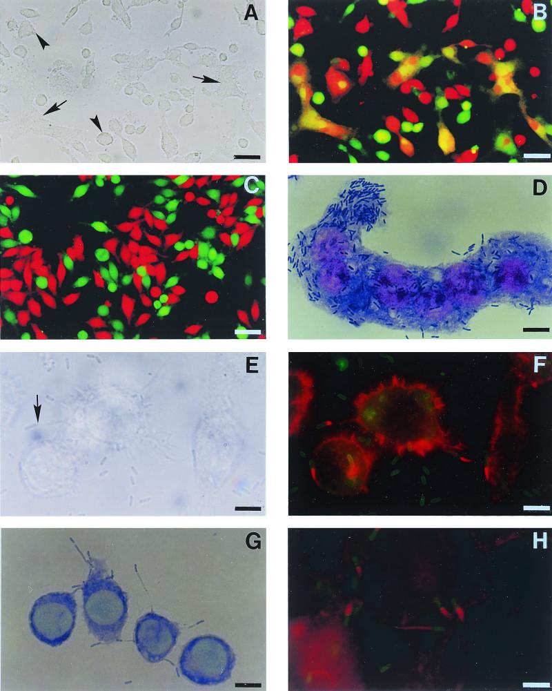
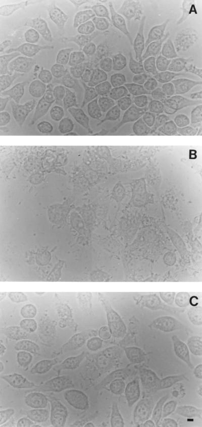
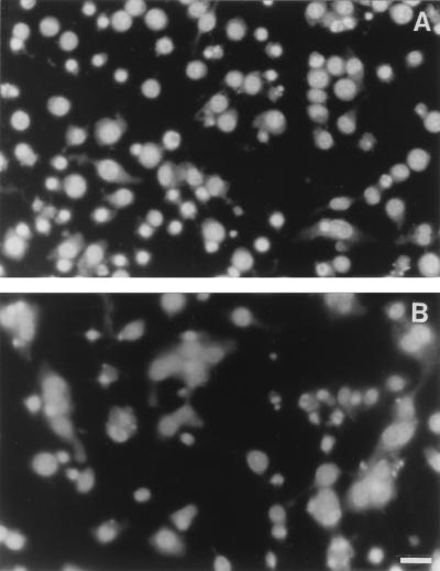
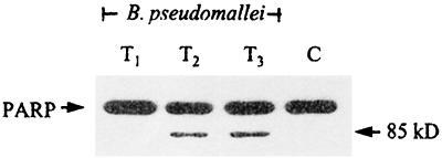
Burkholderia pseudomallei, a facultative intracellular bacterium, is the causative agent of a broad spectrum of diseases collectively known as melioidosis. Its ability to survive inside phagocytic and nonphagocytic cells and to induce multinucleated giant cell (MNGC) formation has been demonstrated. This study was designed to assess a possible mechanism(s) leading to this cellular change, using virulent and nonvirulent strains of B. pseudomallei to infect both phagocytic and nonphagocytic cell lines. We demonstrated that when the cells were labeled with two different cell markers (CMFDA or CMTMR), mixed, and then infected with B. pseudomallei, direct cell-to-cell fusion could be observed, leading to MNGC formation. Staining of the infected cells with rhodamine-conjugated phalloidin indicated that immediately after the infection, actin rearrangement into a comet tail appearance occurred, similar to that described earlier for other bacteria. The latter rearrangement led to the formation of bacterium-containing, actin-associated membrane protrusions which could lead to a direct cell-to-cell spreading of B. pseudomallei in the infected hosts. Results from 4', 6'-diamidine-2-phenylindole dihydrochloride (DAPI) nuclear staining, poly-ADP ribose polymerase cleavage, staining of infected cells for phosphatidylserine exposure with annexin V, and electrophoresis of the DNA extracted from these infected cells showed that B. pseudomallei could kill the host cells by inducing apoptosis in both phagocytic and nonphagocytic cells.


Figures

FIG. 1
FIG. 1
Plaque formation in HeLa cells by B. pseudomallei. Burkholderia plaques occurred in the presence of kanamycin at 24 h after infection. The cell monolayer was stained with neutral red to enhance visibility. Lysis of cells in the center of the plaque could be readily observed (B [magnification, ×40]), leaving at times a considerable amount of visible debris. Cells in the periphery contained a large number of intracellular bacteria.
FIG. 2
FIG. 2
Morphological changes of cells J774A.1 (A through G) and HeLa (H) cells infected with B. pseudomallei. The J774A.1 cells were separately labeled with CMTMR (red) and CMFDA (green) cell markers, mixed, and then cocultured in the presence (A and B) or absence (C) of B. pseudomallei. Cell fusion was observed 6 h later under phase-contrast (A) or fluorescence (B and C) microscopes. Fusion of the differently labeled cells, appearing as orange-staining cells (arrow), could be readily observed (B) in the presence of B. pseudomallei. In the same field (A and B), fusion of the same colored labeling cells can also be seen (arrowhead). In the absence of B. pseudomallei (C), no fusion occurred. An MNGC loaded with numerous bacilli (as indicated by Giemsa stain) could be readily observed at 6 h (D). Phase-contrast (E) and fluorescence (F) photomicrographs demonstrate the presence of actin-based peripheral membrane protrusions (arrow) that occurred 4 h after the infection. The actin tail (red) attached to one pole of the bacterium (green) can be readily observed (F). Contact of the bacterium located at the tip of each protrusion with adjacent cells (as shown by Giemsa stain) is shown in panel G. Similar membrane protrusions with typical actin tails were also noted in nonphagocytic cells (H). Bars = 50 μm (A through C) and 10 μm (D through H).
FIG. 3
FIG. 3
Destruction of J774A.1 cells infected with Ara and Ara+ B. pseudomallei. The cell monolayers (A) were infected with virulent Ara (B) or nonvirulent Ara+ (C) B. pseudomallei for 6 h and then observed under a phase-contrast microscope for MNGC formation and cell destruction. A more extensive morphological change can be readily observed with the virulent organisms (compare panel B with panel C). Bar = 10 μm.
FIG. 4
FIG. 4
Apoptosis of J774A.1 cells infected with B. pseudomallei. The cell monolayer was fixed, and the nuclei were stained with DAPI 4 h (A) and 6 h (B) after the infection. Condensed and fragmented nuclei typical of apoptotic cell death could be readily observed as early as 4 h, when most of the cells were still viable and only a small number of MNGCs had formed. Six hours after the infection, a large number of MNGCs could be readily observed; normal and apoptotic nuclei can appear together within the same MNGC (B). Bar = 50 μm.
FIG. 5
FIG. 5
DNA fragmentation of HeLa cells infected with B. pseudomallei. (A) The cells were infected with a virulent strain of B. pseudomallei (strain 844) and, at the indicated intervals (lanes: 3, 12 h; 4, 14 h; 5, 16 h; 6, 18 h; 7, 20 h; and 8, 24 h), the cells were removed, and the DNA was extracted, subjected to electrophoresis in 1.8% agarose, and stained with ethidium bromide. DNA ladders typical for apoptotic cells could be observed from 18 h of infection onward. Lanes 1 and 2 represent the DNA of uninfected cells from the HeLa line taken at 12 h and 24 h of incubation, respectively. The left lane is the base pair markers. (B) The DNA ladders observed when the cells were infected for 24 h with different strains of B. pseudomallei. Lanes: 2, 3, and 4, virulent strains 844, UE3 and UE12, respectively; 5, 6, and 7, nonvirulent strains UE5, UE8, and UE11, respectively; and 8, 9, and 10, virulent strains, with atypical lipopolysaccharide pattern, UE16, UE30, and 824a, respectively. Lane 1 represents uninfected HeLa cells at 24 h of incubation.
FIG. 6
FIG. 6
PARP cleavage. J774A.1 cells were heavily infected with B. pseudomallei at an MOI of 100:1 for 30 min. After a washing, the infected cells were incubated further for different intervals in the presence of kanamycin, and the cells were then harvested as described in Materials and Methods. Samples removed at 1, 2, and 3 h (lanes T1, T2, and T3, respectively) were lysed and then subjected to immunoblotting. Cleaved PARP (85 kDa) could be readily detected from 2 h (T2) onward. Lane C, Uninfected cell control.

References

    1. Ahmed K, Enciso H D, Masaki H, Tao M, Omori A, Tharavichikul P, Nagatake T. Attachment of Burkholderia pseudomallei to pharyngeal epithelial cells. A highly pathogenic bacteria with low attachment ability. Am J Trop Med Hyg. 1999;60:90–93. - PubMed
    1. Anuntagool N, Intachote P, Wuthiekanun V, White N J, Sirisinha S. Lipopolysaccharide from nonvirulent Ara+Burkholderia pseudomallei isolates is immunologically indistinguishable from lipopolysaccharide from virulent Ara− clinical isolates. Clin Diagn Lab Immunol. 1998;5:225–229. - PMC - PubMed
    1. Chaowagul W, Suputtamongkol Y, Dance D A B, Rachanuvong A, Pattaraarechachai J, White N J. Relapse in melioidosis: incidence and risk factors. J Infect Dis. 1993;168:1181–1185. - PubMed
    1. Chaowagul W, White N J, Dance D A B, Wattanagoon Y, Naigowit P, Davis T M E, Looareesuwan S, Pitakwatchara N. Melioidosis: a major cause of community-acquired septicemia in northeastern Thailand. J Infect Dis. 1989;159:890–899. - PubMed
    1. Cossart P, Boquet P, Normark S, Rappuoli R, editors. Cellular microbiology. Washington, D.C.: ASM Press; 2000. - PubMed

Publication types

LinkOut - more resources