. 2000 Aug;12(8):1257-66.
doi: 10.1105/tpc.12.8.1257.

Prenylation of the floral transcription factor APETALA1 modulates its function

Affiliations

Prenylation of the floral transcription factor APETALA1 modulates its function

S Yalovsky et al. Plant Cell. 2000 Aug.

Abstract

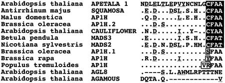
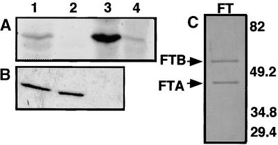
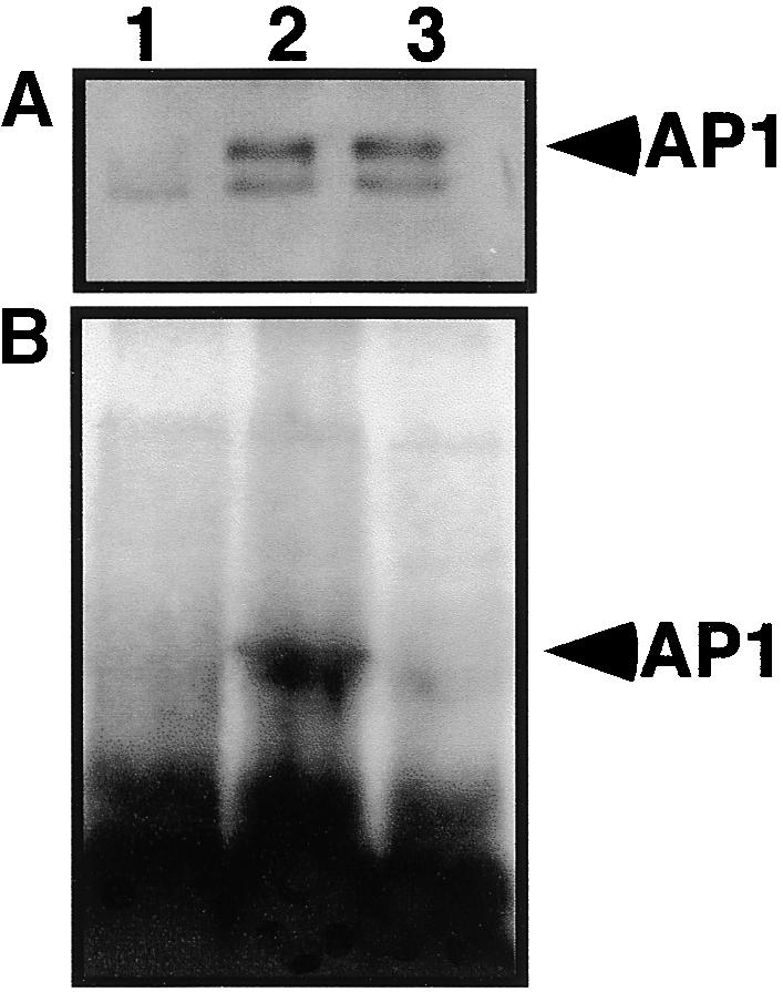
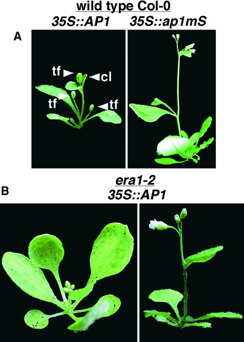
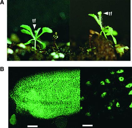
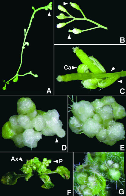
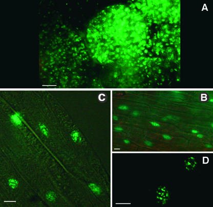
The Arabidopsis MADS box transcription factor APETALA1 (AP1) was identified as a substrate for farnesyltransferase and shown to be farnesylated efficiently both in vitro and in vivo. AP1 regulates the transition from inflorescence shoot to floral meristems and the development of sepals and petals. AP1 fused to green fluorescent protein (GFP) retained transcription factor activity and directed the expected terminal flower phenotype when ectopically expressed in transgenic Arabidopsis. However, ap1mS, a farnesyl cysteine-acceptor mutant of AP1, as well as the GFP-ap1mS fusion protein failed to direct the development of compound terminal flowers but instead induced novel phenotypes when ectopically expressed in Arabidopsis. Similarly, compound terminal flowers did not develop in era1-2 transformants that ectopically expressed AP1. Together, the results demonstrate that AP1 is a target of farnesyltransferase and suggest that farnesylation alters the function and perhaps specificity of the transcription factor.


Figures

Figure 1.
Figure 1.
The CaaX Box Motif Is Conserved between AP1 and Homologs from Distantly Related Plant Species. Amino acid sequence alignment of the C-terminal 20 amino acids of APETALA1 (GenBank accession number Z16421) and selected homologs: SQUAMOSA (X63701) from Antirrhinum majus; AP1H (AJ000759) from Malus domestica (apple); AP1H.2 (U67452) and AP1H.1 (Z37968) from Brassica oleracea; CAULIFLOWER (L36925), AGL8 (Q38876), and AGAMOUS (X53579) from Arabidopsis thaliana; MADS3 (X99653) from Betula pendula; MADS2 (AF068726) from Nicotiana sylvestris; AP1H (AJ251300) from Brassica rapa; and AP1H (AF034093) from Populus tremuloides. The putative CaaX box sequences are shaded; dots indicate residues identical to AP1; dashes indicate gaps formed by the alignment program; the underscoring indicates a conserved amino acid substitution between AP1 and a given homolog; and boxes highlight substitutions of the CaaX box cysteines.
Figure 2.
Figure 2.
In Vitro Prenylation of AP1. (A) GST–AP1 is a protein substrate for prenylation. Reactions in lanes 1 and 3 contained plant FTase extracts from yeast or baculovirus-infected Sf9 cells, respectively, coexpressing the two subunits of tomato FTase. Control extracts were from ram1Δ yeast cells (lane 2) or mock-infected Sf9 cells (lane 4). (B) Farnesylation of GST–AP1 requires a functional CaaX box. Purified plant Ftase, shown in (C), was incubated with GST–AP1 (lane 1), GST–ap1mL (lane 2), and GST–ap1mS (lane 3). (C) Purified recombinant plant FTase prepared from baculovirus-infected Sf9 cells. Positions of protein bands for FTase subunits α and β (FTA and FTB, respectively) are indicated. Numbers at right indicate positions of molecular-weight markers (Bio-Rad) in kilodaltons.
Figure 3.
Figure 3.
AP1 Is Prenylated in Vivo. (A) Immunoblots of protein extracts from PVX-infected N. benthamiana leaves labeled with 3H-mevalonic acid. After infection with control PVX (lane 1), PVX–AP1 (lane 2), or PVX–ap1mS (lane 3) viruses, protein extracts were subjected to immunoblot analysis with polyclonal antibodies prepared against the C-terminal third of AP1. (B) In vivo labeling of AP1. Fluorography of gel with the same protein extracts as shown in (A).
Figure 4.
Figure 4.
Ectopic Expression of AP1 and ap1mS in Wild-Type and era1-2 Plants. (A) Transgenic Arabidopsis Col-0 plants expressing either wild-type AP1 or farnesyl cysteine–acceptor mutant ap1mS under the control of the CaMV 35S promoter (35S::AP1 and 35S::ap1mS, respectively). cl, curled leaf; tf, compound terminal flowers. (B) Transgenic Arabidopsis era1-2 mutant plants expressing wild-type AP1 under control of the CaMV 35S promoter (35S::AP1).
Figure 5.
Figure 5.
A GFP–AP1 Fusion Protein Retains Ectopic AP1 Transcription Factor Activity. (A) Transgenic Arabidopsis Col-0 plants expressing a chimeric gene for the GFP–AP1 fusion protein under the control of the CaMV 35S promoter. tf, compound terminal flowers. (B) Confocal laser scanning microscope images of a gynoecium (left) and a petal area (right) demonstrating the ectopic expression and nuclear localization of GFP–AP1 in transgenic plants shown in (A). formula image; formula image.
Figure 6.
Figure 6.
Ectopic Expression of GFP–ap1mS Did Not Induce Development of Compound Terminal Flowers. Shown are four plants that expressed a high level of the farnesyl cysteine–acceptor mutant ap1mS fused to GFP (GFP–ap1mS) under the control of CaMV 35S promoter. (A) A six-week-old plant with an elongated stem that produced numerous flowers. The primary stem inflorescence (arrowhead) did not terminate in a compound flower. (B) Primary stem inflorescence of the plant shown in (A). Elongated gynoecia, which protruded from closed flowers, are visible (arrowheads). (C) An axillary inflorescence from the plant shown in (A). An elongated stem between the first whorl organs and the rest of the flower (arrowheads) and a carpeloid leaf (Ca) with stigmatic papillae are clearly visible. (D) Primary stem inflorescence composed of numerous colorless flowers and a flower with an elongated gynoecium (arrowhead). (E) Primary stem inflorescence consisting of numerous colorless flowers. Many trichomes are visible. (F) A plant with nonterminal primary (P) and axillary (Ax) inflorescences. (G) The axillary inflorescences from the same plant shown in (F) in more detail. An inflorescence with multiple flowers and the increased number of hair cells on both leaves and sepals are visible.
Figure 7.
Figure 7.
Nuclear Localization of the GFP–ap1mS Fusion Proteins. (A) A flower bud from the plant shown in Figures 6F and 6G. (B) and (C) GFP fluorescence is localized exclusively to nuclei. Images of stamen (B) and sepal (C) tissues were taken from the plant shown in Figures 6A to 6C. Images were produced under dim light and UV illumination (to induce GFP fluorescence) by using Nomarsky differential interference contrast optics to visualize cell boundaries. (D) High-magnification image of two of the nuclei shown in (C). The GFP–ap1mS produces speckles, which are dispersed throughout the nuclear matrix. formula image; formula image.

References

    1. Anant, J.S., Ong, O.C., Xie, H., Clarke, S., O'Brein, P.J., and Fung, B.K.K. (1992). In vivo differential prenylation of retinal cyclic GMP phosphodiesterase catalytic subunits. J. Biol. Chem. 267, 687–690. - PubMed
    1. Anderegg, R.J., Betz, R., Carr, S.A., Crabb, J.W., and Duntze, W. (1988). Structure of the Saccharomyces cerevisiae mating hormone a-factor: Identification of S-farnesyl cysteine as a structural component. J. Biol. Chem. 263, 18236–18240. - PubMed
    1. Bechtold, N., Ellis, J., and Pelletier, G. (1993). In planta Agrobacterium–mediated gene transfer by infiltration of adult Arabidopsis thaliana plants. C. R. Acad. Sci. Ser. III 316, 1194–1199.
    1. Bent, A.F., Kunkel, B.N., Dahlbeck, D., Brown, K.L., Schmidt, R., Giraudat, J., Leung, J., and Staskawicz, B.J. (1994). RPS2 of Arabidopsis thaliana: A leucine-rich repeat class of plant disease resistance genes. Science 265, 1856–1860. - PubMed
    1. Bowman, J.L., Alvarez, J., Weigel, D., Meyerowitz, E.M., and Smyth, D.R. (1993). Control of flower development in Arabidopsis thaliana by APETALA1 and interacting genes. Development 119, 721–743.

Publication types

MeSH terms