. 2000 Aug 15;14(16):1997-2002.

The Dlk1 and Gtl2 genes are linked and reciprocally imprinted

Affiliations

The Dlk1 and Gtl2 genes are linked and reciprocally imprinted

J V Schmidt et al. Genes Dev. .

Abstract

Genes subject to genomic imprinting exist in large chromosomal domains, probably reflecting coordinate regulation of the genes within a cluster. Such regulation has been demonstrated for the H19, Igf2, and Ins2 genes that share a bifunctional imprinting control region. We have identified the Dlk1 gene as a new imprinted gene that is paternally expressed. Furthermore, we show that Dlk1 is tightly linked to the maternally expressed Gtl2 gene. Dlk1 and Gtl2 are coexpressed and respond in a reciprocal manner to loss of DNA methylation. These genes are likely to represent a new example of coordinated imprinting of linked genes.


Figures

Figure 1
Figure 1
Imprinting of the Dlk1 gene in Peromyscus and Mus. (A) Differential display gel showing the polymorphism for Dlk1. (BW) P. maniculatus; (PO) P. polionotus; in the reciprocal crosses the female is listed first. (B) Dlk1 SSCP analysis for imprinting in Peromyscus placenta. RT–PCR was performed using E18.5 placental RNA from the BW and PO parental strains and reciprocal F1 crosses, as well as a 1:1 mixture of BW and PO RNAs (BW + PO). (C) Dlk1 SSCP analysis for imprinting in Peromyscus embryo. RT–PCR was performed using E18.5 embryo RNA from reciprocal F1 crosses. The polymorphism is the same as that shown in B, however the gel was run longer to highlight the difference in migration. (D) Imprinting of Dlk1 in Mus embryo. Direct sequencing was performed on RT–PCR products from E12.5 C57BL/6 (B) and Cast/Ei (C) embryos and F1 offspring (B × C and C × B). The polymorphic base is bold and underlined.
Figure 2
Figure 2
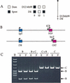
Linkage of the Dlk1 and Gtl2 genes on Mus chromosome 12 and maternal expression of Gtl2. (A) Genotypes of 20 (BTBR × M. spretus) × BTBR backcross animals typed for D12Mit99 and Dlk1. The mapping places Dlk1 1 cm distal to D12Mit99. (B) Genomic organization of Dlk1 and Gtl2 genes compared to H19 and Igf2. Blue boxes denote paternally expressed genes and pink boxes denote maternally expressed genes. The arrows indicate the ICR that regulates imprinting of H19 and Igf2 and the insertion site of the lacZ gene trap vector in the Gtl2lacZ mice (lacZ). (C) RT–PCR analysis for Mus Gtl2 imprinting. RT–PCR products from E12.5 C57BL/6 (B) and Cast/Ei (C) and F1 embryo (E) and placenta (P) were digested with SfcI and analyzed by acrylamide gel electrophoresis.
Figure 3
Figure 3
Coordinate expression of the Dlk1 and Gtl2 genes in Mus embryo and adult. (A) Northern analysis for Dlk1 and Gtl2 RNA in Mus embryos at various stages of development. (B) Northern analysis for Dlk1 and Gtl2 RNA in adult Mus tissues. The lane designations, left to right, are kidney, pituitary, adrenal, brain, testis, and liver. For both blots the expression of the β-actin gene is included as a loading control. The positions of RNA size markers are indicated.
Figure 4
Figure 4
Altered expression of the Dlk1 and Gtl2 genes in Dnmt−/− (Dnmts) embryos. (A) Hybridization of wild-type (WT), Dnmt−/−, and Dnmt−/−/H19Δ13 RNA to the Atlas cDNA expression microarray containing the genes indicated. (B) Northern analysis for Dlk1 and Gtl2 expression in wild-type and Dnmt−/−/H19Δ13 embryos. Expression of the rpL32 gene is included as a loading control.
Figure 5
Figure 5
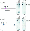
Methylation analysis of the Gtl2 and Dlk1 promoter regions. (A) Methylation analysis of the Gtl2 promoter and first exon by Southern blotting using HincII (H), HincII + MspI (H + M) or HincII + HpaII (H + Hp). The location of the MspI/HpaII sites are indicated by the arrows and the probe by the dark line. Molecular DNA markers are indicated to the right of the gel. (B) Methylation analysis of the Dlk1 promoter and first exon. Abbreviations as in A.

References

    1. Bachmann E, Krogh TN, Hojrup P, Skjodt K, Teisner B. Mouse fetal antigen 1 (mFA1), the circulating gene product of mdlk, pref-1 and SCP-1: Isolation, characterization and biology. J Reprod Fertil. 1996;107:279–285. - PubMed
    1. Barlow DP, Stoger R, Herrmann BG, Saito K, Schweifer N. The mouse insulin-like growth factor type-2 receptor is imprinted and closely linked to the Tme locus. Nature. 1991;349:84–87. - PubMed
    1. Barlow DP. Competition—a common motif for the imprinting mechanism? EMBO J. 1997;16:6899–6905. - PMC - PubMed
    1. Bauer SR, Ruiz-Hidalgo MJ, Rudikoff EK, Goldstein J, Laborda J. Modulated expression of the epidermal growth factor-like homeotic protein dlk influences stromal-cell-pre-B-cell interactions, stromal cell adipogenesis, and pre-B-cell interleukin-7 requirements. Mol Cell Biol. 1998;18:5247–5255. - PMC - PubMed
    1. Bell AC, Felsenfeld G. Methylation of a CTCF-dependent boundary controls imprinted expression of the Igf2 gene. Nature. 2000;405:482–485. - PubMed

Publication types

LinkOut - more resources