. 2000 Aug 15;14(16):2046-59.

The checkpoint protein Ddc2, functionally related to S. pombe Rad26, interacts with Mec1 and is regulated by Mec1-dependent phosphorylation in budding yeast

Affiliations

The checkpoint protein Ddc2, functionally related to S. pombe Rad26, interacts with Mec1 and is regulated by Mec1-dependent phosphorylation in budding yeast

V Paciotti et al. Genes Dev. .

Abstract

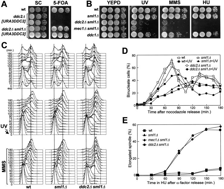
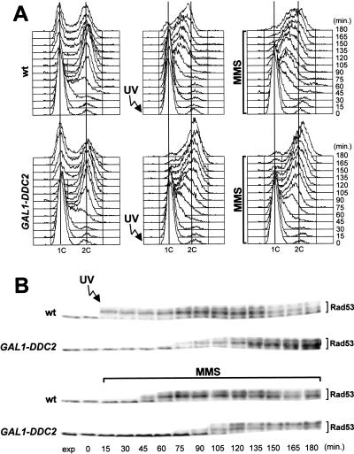
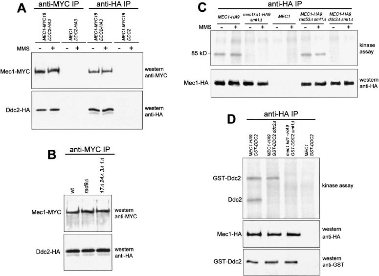
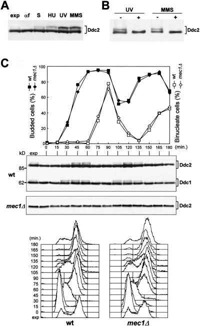
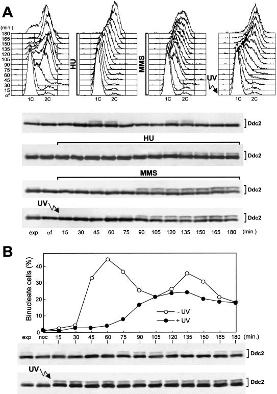
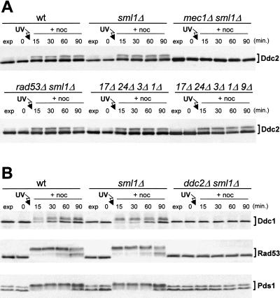
DDC2 is a novel component of the DNA integrity checkpoint pathway, which is required for proper checkpoint response to DNA damage and to incomplete DNA replication. Moreover, Ddc2 overproduction causes sensitivity to DNA-damaging agents and checkpoint defects. Ddc2 physically interacts with Mec1 and undergoes Mec1-dependent phosphorylation both in vitro and in vivo. The phosphorylation of Ddc2 takes place in late S phase and in G(2) phase during an unperturbed cell cycle and is further increased in response to DNA damage. Because Ddc2 phosphorylation does not require any other known tested checkpoint factors but Mec1, the Ddc2-Mec1 complex might respond to the presence of some DNA structures independently of the other known checkpoint proteins. Our findings suggest that Ddc2 may be the functional homolog of Schizosaccharomyces pombe Rad26, strengthening the hypothesis that the mechanisms leading to checkpoint activation are conserved throughout evolution.


Figures

Figure 1
Figure 1
DNA damage hypersensitivity and checkpoint defects of ddc2Δ sml1Δ cells. Strains were as follows: wild type (K699), ddc2Δ [URA3 DDC2] (YLL275), ddc2Δ sml1Δ [URA3 DDC2] (DMP2995/7A), sml1Δ (YLL488) (Longhese et al. 2000), ddc2Δ sml1Δ (DMP2995/1B), mec1Δ sml1Δ (YLL490) (Longhese et al. 2000), and ddc1Δ (YLL244). (A–B) Serial dilution of YEPD-exponentially growing cell cultures were spotted on SC plates with or without 5-FOA (A) or on YEPD plates with or without MMS (0.005%) or HU (5 mm) (B). YEPD plates were made in duplicate, and one of them was UV-irradiated (30 J/m2) (UV). (C) α-Factor-synchronized cells were released from α-factor at time zero in YEPD (top), were UV-irradiated (40 J/m2) prior to the release in YEPD (middle), or were released in YEPD containing 0.02% MMS (bottom). Samples of untreated, UV- and MMS-treated cell cultures were collected at the indicated times after α-factor release and analyzed by FACS. (D) Cell cultures were arrested with nocodazole and were UV-irradiated (50 J/m2). Cell cycle progression was monitored at the indicated times in unirradiated and UV-irradiated cultures after release from nocodazole, by direct visualization of nuclear division by propidium iodide staining. (E) Cell cultures were arrested in G1 with α-factor and then released at time zero in YEPD containing 200 mm HU. Aliquots of cells were collected at the indicated times and stained with antitubulin antibodies to score for the percentage of cells with elongated spindles by indirect immunofluorescence. FACS analysis of the DNA content and plating for cell survival were carried out concomitantly (see text for details).
Figure 2
Figure 2
Overexpression of DDC2 causes DNA damage checkpoint defects. Cultures of wild type (K699) and GAL1–DDC2 (YLL279.4) strains, logarithmically growing in YEP-raffinose, were synchronized with α-factor. Galactose to 2% was added 2.5 hr before α-factor addition. Release from α-factor block was performed by transferring cell cultures to YEP medium containing both raffinose and galactose, with or without 0.02% MMS. One-third of each α-factor-synchronized culture was UV-irradiated prior to the release in YEP–raf–gal. Time zero corresponds to cell samples withdrawn immediately before MMS addition or UV-irradiation and release from α-factor. The data presented in panels A and B all come from the same experiment. (A) Samples of untreated (left), UV-irradiated (middle), or MMS-treated (right) cells were taken at the indicated times after α-factor release and analyzed by FACS. (B) Extracts from UV- or MMS-treated cell cultures were analyzed by Western blot with anti-Rad53 antibodies. (exp) Exponentially growing cells.
Figure 3
Figure 3
Physical interaction between Ddc2 and Mec1 and in vitro kinase assay. (A) Immunoprecipitations with anti-MYC (anti-MYC IP) or anti-HA (anti-HA IP) antibodies were performed on extracts from exponentially growing untreated (−) or MMS-treated (+) (0.02% MMS for 1 hr) cells expressing Mec1–MYC18 (YLL447.32/1A) or Ddc2–HA3 (YLL683.8/3B) or both (DMP3084/3C), as indicated in the top part of the panel. Mec1 and Ddc2 were then detected by Western blot analysis of the immunoprecipitates probed with the antibodies indicated on the right side of the panel. (B) Western blot analysis of the anti-MYC immunoprecipitates of protein extracts from untreated wild-type (DMP3084/3C), rad9Δ (DMP3167/18A), and rad17Δ rad24Δ mec3Δ ddc1Δ (DMP3168/6D) cells concomitantly expressing Ddc2–HA3 and Mec1–MYC18. (C–D) Kinase assays were performed on anti-HA immunoprecipitates of protein extracts from exponentially growing untreated (−) or MMS-treated (+) cells with the genotypes indicated in the top parts of the panels. The same immunoprecipitates were also analyzed by Western blot using the antibodies indicated in the bottom parts of the right side of the panels. (C) Strains MEC1–HA9 (YLL476.34/2C), mec1kd1–HA9 sml1Δ (YLL593.1.3), MEC1 (K699), MEC1–HA9 rad53Δ sml1Δ (DMP2959/3A), MEC1–HA9 ddc2Δ sml1Δ (DMP3014/2A). (D) Strains MEC1–HA9 [pML103 GAL1–GST–DDC2] (YLL680), MEC1–HA9 ddc2Δ [pML103 GAL1–GST–DDC2] (YLL681), mec1kd1–HA9 sml1Δ [pML103 GAL1–GST–DDC2] (YLL682), and MEC1 [pML103 GAL1–GST–DDC2] (YLL678).
Figure 4
Figure 4
Ddc2 is phosphorylated during an unperturbed cell cycle and in response to DNA insults. (A–B) Protein extracts were prepared from strain YLL683.8/3B, expressing Ddc2–HA3 from the DDC2 promoter. (A) Western blot analysis with anti-HA antibodies of protein extracts from exponentially growing cultures untreated (exp) or treated with HU (50 mm; 1 hr), UV (40 J/m2), or MMS (0.02%; 1 hr). Protein extracts were also prepared from G1-arrested cells (αf) or from cells progressing through S phase 30 min after release from α-factor (S). (B) Protein extracts from UV- and MMS-treated exponentially growing cells were immunoprecipitated with anti-HA antibodies. Immunoprecipitates were then incubated at 30°C with (+) or without (−) λ-phosphatase, before electrophoresis and Western blot analysis using anti-HA antibodies. (C) Exponentially growing wild-type (DMP3198/1A) cells, concomitantly expressing Ddc2–HA3 and Ddc1–HA2 from the corresponding promoters, and mec1Δ sml1Δ (DMP3048/5B) cells, expressing Ddc2–HA3, were synchronized with α-factor and released into the cell cycle at time zero. Samples were collected at the indicated times after α-factor release to determine the percentage of budded and binucleate cells (top) and to perform Western blot analysis with anti-HA antibodies of protein extracts (middle) (molecular mass markers, kD, are indicated) and FACS analysis of the DNA content (bottom).
Figure 5
Figure 5
Ddc2 phosphorylation in response to DNA damage in different cell cycle phases or to replication block. The strain is YLL683.8/3B, expressing Ddc2–HA3 from the DDC2 promoter. (A) Cell cultures were synchronized with α-factor (αf) and released from the pheromone block in YEPD, YEPD containing 50 mm HU, YEPD containing 0.02% MMS, or were UV-irradiated (40 J/m2) prior to the release in YEPD. Cell samples collected at the indicated times after α-factor release were analyzed by FACS, and protein extracts were prepared and analyzed by Western blot with anti-HA antibodies. (B) Cell cultures were arrested with nocodazole (noc) and UV-irradiated (50 J/m2) prior to the release from the nocodazole arrest. Cell samples of unirradiated and UV-irradiated cultures collected at the indicated times after nocodazole release were analyzed for the percentage of nuclei division, and protein extracts were prepared and analyzed by Western blot using anti-HA antibodies.
Figure 6
Figure 6
Interdependency of phosphorylation events in the checkpoint response. Cell cultures were arrested with nocodazole, UV-irradiated (50 J/m2), and resuspended in YEPD containing 15 μg/ml nocodazole (+noc). Time zero corresponds to cell samples taken immediately before UV irradiation. (A) Western blot analysis of protein extracts from wild-type (YLL683.8/3B), sml1Δ (DMP3048/8B), mec1Δ sml1Δ (DMP3048/5B), rad53Δ sml1Δ (DMP3080/7A), ddc1Δ rad24Δ rad17Δ mec3Δ (DMP3145/2D), and ddc1Δ rad24Δ rad17Δ mec3Δ rad9Δ (DMP3146/4B) strains, expressing Ddc2–HA3 from the DDC2 promoter. In all panels the Ddc2–HA3 protein was visualized with anti-HA antibodies on Western blots of protein extracts prepared at the indicated times. (B) The top part of the panel shows Western blot analysis with anti-HA monoclonal antibodies of protein extracts from wild-type (DMP3172/3A), sml1Δ (DMP3172/8A), and ddc2Δ sml1Δ (DMP3172/8C) cells, expressing Ddc1–HA2 from the DDC1 promoter. The middle and bottom parts of the panel show Western blot analysis with anti-Rad53 polyclonal antibodies and anti-MYC monoclonal antibodies, respectively, of protein extracts from wild-type (DMP3171/2A), sml1Δ (DMP3171/8B), and ddc2Δ sml1Δ (DMP3171/8A) cells, expressing Pds1–MYC18 from the PDS1 promoter.

References

    1. Bashkirov VI, King JS, Bashkirova EV, Schmuckli-Maurer J, Heyer WD. DNA repair protein Rad55 is a terminal substrate of the DNA damage checkpoints. Mol Cell Biol. 2000;20:4393–4404. - PMC - PubMed
    1. Bentley NJ, Holtzman DA, Flaggs G, Keegan KS, Demaggio A, Ford JC, Hoekstra M, Carr AM. The Schizosaccharomyces pombe rad3 checkpoint gene. EMBO J. 1996;15:6641–6651. - PMC - PubMed
    1. Carr AM. Control of cell cycle arrest by the Mec1sc/Rad3sp DNA structure checkpoint pathway. Curr Opin Genet Dev. 1997;7:93–98. - PubMed
    1. Caspari T, Dahlen M, Kanter-Smoler G, Lindsay HD, Hofmann K, Papadimitriou K, Sunnerhagen P, Carr AM. Characterization of Schizosaccharomyces pombe Hus1: A PCNA-related protein that associates with Rad1 and Rad9. Mol Cell Biol. 2000;74:1254–1262. - PMC - PubMed
    1. Chen G, Yuan SF, Liu W, Xu Y, Trujillo K, Song B, Cong F, Goff SP, Wu Y, Arlinghaus R, et al. Radiation-induced assembly of Rad51 and Rad42 recombination complex requires ATM and c-Abl. J Biol Chem. 1999;274:12748–12752. - PubMed

Publication types

MeSH terms