. 2000 Aug 15;14(16):2060-71.

tBID, a membrane-targeted death ligand, oligomerizes BAK to release cytochrome c

Affiliations

tBID, a membrane-targeted death ligand, oligomerizes BAK to release cytochrome c

M C Wei et al. Genes Dev. .

Abstract

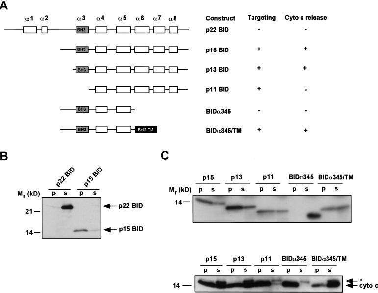
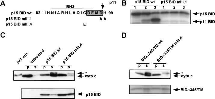
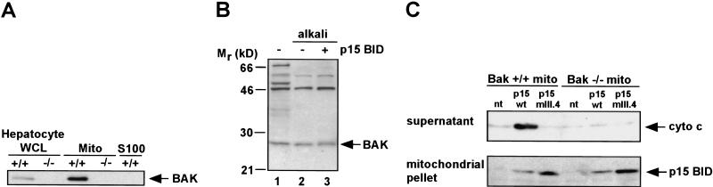
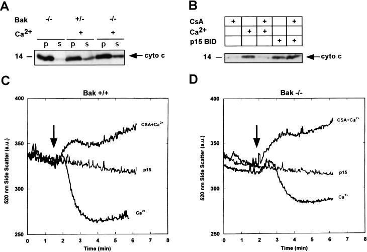
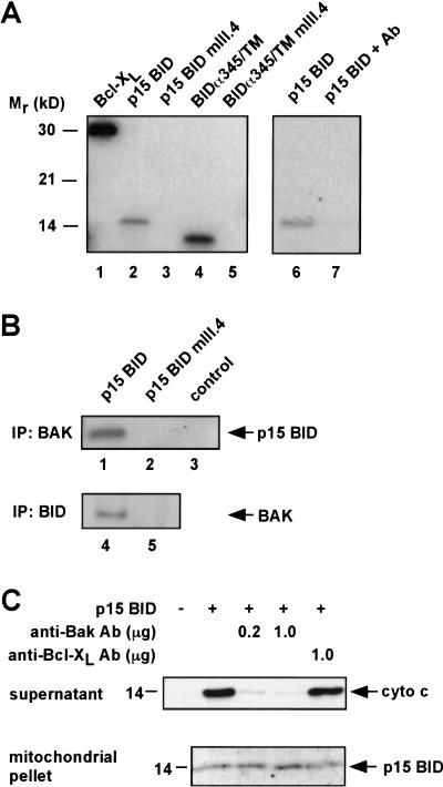
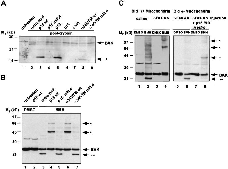
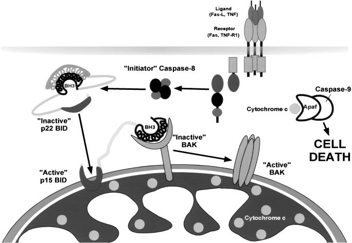
TNFR1/Fas engagement results in the cleavage of cytosolic BID to truncated tBID, which translocates to mitochondria. Immunodepletion and gene disruption indicate BID is required for cytochrome c release. Surprisingly, the three-dimensional structure of this BH3 domain-only molecule revealed two hydrophobic alpha-helices suggesting tBID itself might be a pore-forming protein. Instead, we demonstrate that tBID functions as a membrane-targeted death ligand in which an intact BH3 domain is required for cytochrome c release, but not for targeting. Bak-deficient mitochondria and blocking antibodies reveal tBID binds to its mitochondrial partner BAK to release cytochrome c, a process independent of permeability transition. Activated tBID results in an allosteric activation of BAK, inducing its intramembranous oligomerization into a proposed pore for cytochrome c efflux, integrating the pathway from death receptors to cell demise.


Figures

Figure 1
Figure 1
In vitro targeting and cytochrome c release by various BID proteins. IVT proteins were incubated with purified mitochondria. The mitochondria were centrifuged, and pellet and supernatant were assayed for targeting and cytochrome c release. (A) Schematic of BID constructs showing α helices and capacity to target mitochondria and release cytochrome c. (B) Anti-BID Western blot of an in vitro targeting assay of p22 and p15 BID. Mitochondrial pellet (p) and supernatant (s). (C) Autoradiogram of 35S-labeled IVT BID proteins assayed by in vitro targeting (top). Anti-cytochrome c Western blot assessing cytochrome c release from pellet (p) into supernatant (s) (bottom). (*) A cross-reactive protein present in IVT mix.
Figure 2
Figure 2
Requirement for an intact BH3 domain on the cytoplasmic face of the mitochondrial outer membrane. (A) Sequence of p15 BID BH3 domain mutants mIII.1 and mIII.4. Shaded box indicates the caspase-3 cleavage site DEMD that generates the p11 fragment. (B) Treatment of mitochondria with recombinant caspase-3 after targeting wild-type (wt) and caspase-cleavage site mutant p15 BID mIII.1. (Lane 1) Amount of p15 BID targeted. (Lane 2) Treatment with caspase-3 at 4°C. (Lane 3) Treatment with caspase-3 at 30°C. p15 BID and p11 BID are visualized by 35S fluorography. (C) In vitro targeting and cytochrome c release by p15 BID wild type vs mIII.4. (*) A cross-reactive protein present in IVT mix. (D) In vitro targeting and cytochrome c release by BIDα345/TM wild-type and mutant mIII.4.
Figure 3
Figure 3
Requirement for BAK in p15 BID-induced cytochrome c release. (A) Subcellular fractionation of Bak-deficient (−/−) and wild-type ( +/+) hepatocytes, immunoblotted with an anti-BAK Ab. (WCL) Whole cell lysate, (Mito) heavy membrane, (S100) cytosol fraction. Equal amounts of protein were loaded in the paired WCL and Mito fractions. (B) Alkali-resistance of mitochondrial BAK in the presence or absence of p15 BID. Wild-type mitochondria +/− 0.5 ng/μl recombinant p15 BID were resuspended in 0.1 m Na2CO3 (pH 11.5) at 4°C for 30 min, and the mitochondrial membranes were then pelleted and analyzed by an anti-BAK Western blot. (C) In vitro targeting and cytochrome c release by recombinant p15 BID wild-type and mutant p15 mIII.4 in either Bak-deficient (−/−) or wild-type (+/+) mitochondria.
Figure 4
Figure 4
p15 BID-induced cytochrome c release is independent of permeability transition (PT), and BAK is not required for PT. (A) Cytochrome c release in response to 200 μm Ca2+, which induces permeability transition in Bak-deficient (−/−) and wild-type or heterozygous (+/−) isolated mitochondria. (B) Comparison of Ca2+ vs p15 BID-induced cytochrome c release and response to 1 μm cyclosporin A (CsA) in isolated mitochondria. (C,D) Mitochondrial swelling as determined by a real time measure of side scatter in response to 75 μm Ca2+, or 75 μm Ca2+ plus 1.5 μm CsA, or p15 BID (0.5 ng/μl) in Bak+/+ (C) or Bak−/− isolated mitochondria (18 μg mitochondrial protein in 2 ml of targeting buffer). (D) Arrows indicate time of p15 BID or Ca2+ addition.
Figure 5
Figure 5
BID and BAK physically interact, and a BAK-blocking Ab prevents cytochrome c release. (A) In vitro binding between GST–BAK and IVT BCL-XL, p15 BID, and BIDα345/TM, but not p15 BID mIII.4 or BIDα345/TM mIII.4 (lanes 1–5). None of the IVT proteins bound GST itself (data not shown). Preincubation with an anti-BAK Ab inhibits GST–-BAK in vitro binding to p15 BID (lanes 6,7). (B) Coimmunoprecipitation of BAK and wild-type p15 BID. 35S-labeled p15 BID wild-type (lane 1) and mutant p15 BID mIII.4 (lane 2) were targeted to mitochondria in vitro. The mitochondria were then solubilized and immunoprecipitated with an anti-BAK Ab. Coimmunoprecipitated p15 was detected by autoradiography. The anti-BAK Ab did not directly immunoprecipitate p15 BID wild type from solution (lane 3). BAK (24 kD) comigrates with light chain (25 kD), precluding detection of BAK by IP Western. Consequently, 35S-labeled IVT BAK was mixed with IVT p15 wild type or p15 mIII.4 in buffer B. BID was immunoprecipitated with an anti-BID Ab, and coimmunoprecipitated BAK was detected by autoradiography (lanes 4,5). (C) Inhibition of the p15 BID/BAK interaction prevents cytochrome c release. Wild-type mitochondria were incubated with the indicated amounts of anti-BAK Ab (G-23) or anti-BCL-XL Ab (SC-18) for 20 min at room temperature. A total of 25 ng of recombinant p15 BID was targeted to the mitochondria. The cytochrome c released into the supernatant and the p15 BID targeted to the mitochondrial pellet are shown.
Figure 6
Figure 6
p15 BID induces a conformational change and oligomerization of BAK in vitro and in vivo. (A) The IVT BID proteins denoted were targeted to wild-type mitochondria. The mitochondria were then treated with 30 μg/ml trypsin (lanes 2–9). The trypsin sensitivity pattern of BAK was assessed by an anti-BAK Ab Western blot. (B) The IVT BID proteins denoted were targeted to mitochondria. Mitochondria were then treated with a DMSO control buffer or with 10 mm BMH cross-linker. Pattern of cross-linked BAK was determined by an anti-BAK Ab Western blot. (*) Higher molecular mass BMH-cross-linked BAK complexes. (**) An intramolecularly cross-linked BAK monomer displaying faster mobility. (C) Mitochondria were compared from Bid+/+ versus Bid−/− mice following intravenous injection of saline or anti-Fas Ab (Jo2, Pharmingen, 0.5 μg/gm body mass) for 2 hr. Fas-activated Bid+/+ mitochondria lost the faster mobility BAK species and demonstrated BMH cross-linked higher molecular mass BAK complexes (lanes 3,4). Bid−/− mitochondria do not oligomerize BAK in response to in vivo Fas activation (lanes 5,6). However, BAK oligomerization can be restored by in vitro targeting of p15 BID (lanes 7,8).
Figure 7
Figure 7
An activation cascade of pro-apoptotic BID to BAK integrates the apoptotic pathway from death receptors to mitochondrial release of cytochrome c.

References

    1. Adams JM, Cory S. The Bcl-2 protein family: Arbiters of cell survival. Science. 1998;281:1322–1326. - PubMed
    1. Antonsson B, Conti F, Ciavatta A, Montessuit S, Lewis S, Martinou I, Bernasconi L, Bernard A, Mermod JJ, Mazzei G, et al. Inhibition of Bax channel-forming activity by Bcl-2. Science. 1997;277:370–372. - PubMed
    1. Basanez G, Nechushtan A, Drozhinin O, Chanturiya A, Choe E, Tutt S, Wood KA, Hsu Y, Zimmerberg J, Youle RJ. Bax, but not Bcl-xL, decreases the lifetime of planar phospholipid bilayer membranes at subnanomolar concentrations. Proc Natl Acad Sci. 1999;96:5492–5497. - PMC - PubMed
    1. Cecconi F, Alvarez-Bolado G, Meyer BI, Roth KA, Gruss P. Apaf1 (CED-4 homolog) regulates programmed cell death in mammalian development. Cell. 1998;94:727–737. - PubMed
    1. Chou JJ, Li H, Salvesen GS, Yuan J, Wagner G. Solution structure of BID, an intracellular amplifier of apoptotic signaling. Cell. 1999;96:615–624. - PubMed

Publication types