. 2000 Aug 15;14(16):2072-84.

Hypersensitivity to DNA damage leads to increased apoptosis during early mouse development

Affiliations

Hypersensitivity to DNA damage leads to increased apoptosis during early mouse development

B S Heyer et al. Genes Dev. .

Abstract

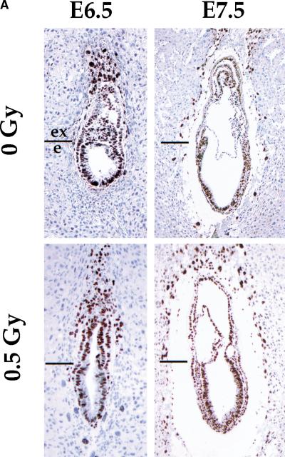
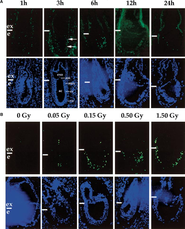
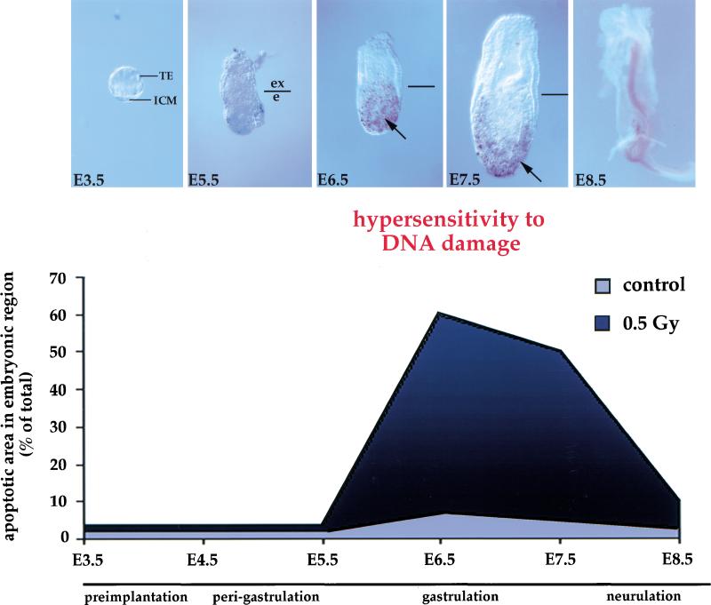
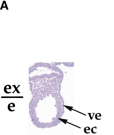
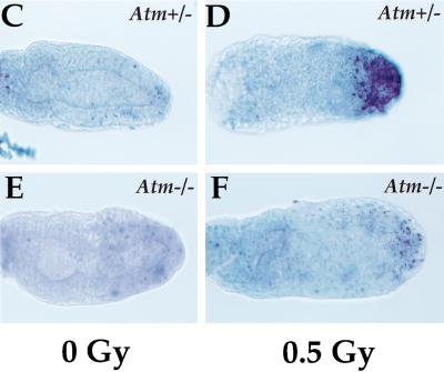
Gastrulation in mice is associated with the start of extreme proliferation and differentiation. The potential cost to the embryo of a very rapid proliferation rate is a high production of damaged cells. We demonstrate a novel surveillance mechanism for the elimination of cells damaged by ionizing radiation during mouse gastrulation. During this restricted developmental window, the embryo becomes hypersensitive to DNA damage induced by low dose irradiation (<0.5 Gy) and undergoes apoptosis without cell cycle arrest. Intriguingly, embryonic cells, including germ cell progenitors, but not extraembryonic cells, become hypersensitive to genotoxic stress and undergo Atm- and p53-dependent apoptosis. Thus, hypersensitivity to apoptosis in the early mouse embryo is a cell fate-dependent mechanism to ensure genomic integrity during a period of extreme proliferation and differentiation.


Figures

Figure 1
Figure 1
Cellular proliferation is not impaired in irradiated embryos. (A) BrdU labeling of E6.5 and E7.5 embryos after low dose irradiation (0.5 Gy). Strongly BrdU-positive nuclei (brown) can be seen throughout the control embryos and irradiated embryos. The boundary between the extraembryonic (ex) and embryonic (e) regions is indicated. (B) Percentage of total number of cells that are BrdU labeled. At E6.5, 70.3 ± 2.9% and 67 ± 1.3% of cells were strongly BrdU positive for the controls (n = 5) and irradiated embryos (n = 4), respectively. At E7.5, 73.25 ± 4.4% and 72.8 ± 5.6% of the nuclei were labeled in the controls (n = 4) and irradiated embryos (n = 7), respectively.
Figure 1
Figure 1
Cellular proliferation is not impaired in irradiated embryos. (A) BrdU labeling of E6.5 and E7.5 embryos after low dose irradiation (0.5 Gy). Strongly BrdU-positive nuclei (brown) can be seen throughout the control embryos and irradiated embryos. The boundary between the extraembryonic (ex) and embryonic (e) regions is indicated. (B) Percentage of total number of cells that are BrdU labeled. At E6.5, 70.3 ± 2.9% and 67 ± 1.3% of cells were strongly BrdU positive for the controls (n = 5) and irradiated embryos (n = 4), respectively. At E7.5, 73.25 ± 4.4% and 72.8 ± 5.6% of the nuclei were labeled in the controls (n = 4) and irradiated embryos (n = 7), respectively.
Figure 2
Figure 2
Time and dose dependence of apoptosis in response to low dose irradiation. (A) E6.5 embryos were exposed in utero to 0.5 Gy and collected at different time points from 1 to 24 hr post-irradiation as indicated. (Top) Embryos probed for apoptotic cells (green); (bottom) embryos stained with Hoechst dye (nuclei detection). (Arrows) Apoptotic embryonic cells. (ac) Amniotic cavity; (am) amnion; (ec) embryonic ectoderm; (exo) exocoelom; (mes) mesoderm, (ve) visceral endoderm. (B) E6.5 embryos were exposed to increasing doses of X-ray from 0 to 1.5 Gy as indicated and collected 6 hr post-irradiation. (Top) Embryos probed for apoptotic cells (green); (bottom) embryos stained with Hoechst dye. (Horizontal white bars) Boundary between the extraembryonic (ex) and embryonic (e) regions.
Figure 3
Figure 3
Apoptosis in response to low dose irradiation (0.5 Gy) is timed with the onset of gastrulation. Embryos were exposed in utero to 0.5 Gy at different time points during development (from E3.5 to E8.5) and collected 6 hr post-irradiation. For each time point, at least 20 embryos were analyzed. The staining in the center of E8.5 embryo is background. The embryos were subsequently probed for apoptotic cells. (Arrows) Apoptotic cells. (Bottom) Graphic representation of the change in hypersensitivity/apoptosis in response to low dose irradiation during early mouse development.
Figure 4
Figure 4
Gastrulating embryos derived from blastocyst outgrowths in culture acquire hypersensitivity to low dose irradiation. (A) Sagittal section of a 4-day outgrowth. The boundary between the extraembryonic (ex) and embryonic (e) regions is indicated. (B) Brachyury expression in blastocyst outgrowths after 2 and 4 days in culture. RT–PCR was performed with Brachyury-specific primers. GAPDH was amplified as a control. (C) Blastocysts were collected at E3.5 and individually cultured with or without EGF. (Top) Phase contrast image; (bottom) apoptotic cells (white). (Arrows) Apoptotic cells. In 2-day outgrowths, apoptosis is induced in the absence of EGF. In 4-day outgrowths, apoptosis is not induced in the absence of EGF. (D) Blastocysts were collected at E3.5 and individually cultured in the presence of EGF; half were irradiated at day 2 or 4 of outgrowth with 0.5 Gy. Irradiation with 0.5 Gy induced apoptosis in 4-day outgrowths but not in 2-day outgrowths. Addition of ZVAD-FMK reduced basal apoptosis in 4-day outgrowths, but did not have an effect on apoptosis induced by X-irradiation. (Open bars) Controls; (black bars) 0.5 Gy irradiation; (gray bars) ZVAD-FMK added. (ec) Embryonic ectoderm; (GC) giant cell; (outg) outgrowth; (ve) visceral endoderm.
Figure 4
Figure 4
Gastrulating embryos derived from blastocyst outgrowths in culture acquire hypersensitivity to low dose irradiation. (A) Sagittal section of a 4-day outgrowth. The boundary between the extraembryonic (ex) and embryonic (e) regions is indicated. (B) Brachyury expression in blastocyst outgrowths after 2 and 4 days in culture. RT–PCR was performed with Brachyury-specific primers. GAPDH was amplified as a control. (C) Blastocysts were collected at E3.5 and individually cultured with or without EGF. (Top) Phase contrast image; (bottom) apoptotic cells (white). (Arrows) Apoptotic cells. In 2-day outgrowths, apoptosis is induced in the absence of EGF. In 4-day outgrowths, apoptosis is not induced in the absence of EGF. (D) Blastocysts were collected at E3.5 and individually cultured in the presence of EGF; half were irradiated at day 2 or 4 of outgrowth with 0.5 Gy. Irradiation with 0.5 Gy induced apoptosis in 4-day outgrowths but not in 2-day outgrowths. Addition of ZVAD-FMK reduced basal apoptosis in 4-day outgrowths, but did not have an effect on apoptosis induced by X-irradiation. (Open bars) Controls; (black bars) 0.5 Gy irradiation; (gray bars) ZVAD-FMK added. (ec) Embryonic ectoderm; (GC) giant cell; (outg) outgrowth; (ve) visceral endoderm.
Figure 5
Figure 5
Irradiation of embryos reduces the number of PGCs. (A) Schematic representation of the localization of PGCs. At E6.5, epiblast cells that become directed to the PGC fate are located in the proximal region of the embryo close to the embryonic/extraembryonic boundary (shaded area). At E8.5, PGCs are located at the base of the allantois around the hindgut pocket (Arrow shows the view for B–D). (B –D). Embryos were stained with alkaline phosphatase (blue) to mark PGCs. (B) Control; (C,D) embryos irradiated with 0.5 Gy at E6.25 or E7.25, respectively, and collected at E8.5. (Arrows) Stained PGCs. (e) Embryonic region; (ex) extraembryonic region; (D) distal; (Pr) proximal.
Figure 6
Figure 6
p53 is a key regulator in the embryonic response to low dose irradiation. (A) E6.5 p53+/+ control; (B) E6.5 p53+/+, 0.5 Gy irradiated; (C) E6.5 p53−/− control; (D) E6.5 p53−/−, 0.5 Gy irradiated. In p53-null mutants the endogenous apoptosis is reduced. In response to low dose irradiation (0.5 Gy), fewer apoptotic cells are seen in the p53-null embryo (D) than in the wild-type littermate (B). (E) The number of apoptotic cells in embryos from p53 heterozygous matings. (Open bars) Controls; (black bars) 0.5 Gy irradiated. The pooled data show that the number of apoptotic cells in response to low dose irradiation is reduced in the absence of p53 (P < 0.001).
Figure 7
Figure 7
Atm mediates apoptosis in response to low dose irradiation in the early embryo. (A) Atm and p53 expression in embryonic and extraembryonic regions at E7 after low dose irradiation (0.5 Gy). RT–PCR was performed with Atm- and p53-specific primers. (B) p53 expression in Atm heterozygous and homozygous null embryos at E7 after low dose irradiation (0.5 Gy). RT–PCR was performed with p53-specific primers. GAPDH was amplified as control in A and B. (C) E6.5 Atm+/− control. (D) E6.5 Atm+/−, 0.5 Gy irradiated. (E) E6.5 Atm−/− control. (F) E6.5 Atm −/−, 0.5 Gy irradiated. In response to low dose irradiation, fewer apoptotic cells are seen in the Atm -null embryo (F) than in the heterozygous littermate (D).
Figure 7
Figure 7
Atm mediates apoptosis in response to low dose irradiation in the early embryo. (A) Atm and p53 expression in embryonic and extraembryonic regions at E7 after low dose irradiation (0.5 Gy). RT–PCR was performed with Atm- and p53-specific primers. (B) p53 expression in Atm heterozygous and homozygous null embryos at E7 after low dose irradiation (0.5 Gy). RT–PCR was performed with p53-specific primers. GAPDH was amplified as control in A and B. (C) E6.5 Atm+/− control. (D) E6.5 Atm+/−, 0.5 Gy irradiated. (E) E6.5 Atm−/− control. (F) E6.5 Atm −/−, 0.5 Gy irradiated. In response to low dose irradiation, fewer apoptotic cells are seen in the Atm -null embryo (F) than in the heterozygous littermate (D).

References

    1. Aladjem MI, Spike BT, Rodewald LW, Hope TJ, Klemm M, Jaenisch R, Wahl GM. ES cells do not activate p53-dependent stress responses and undergo p53-independent apoptosis in response to DNA damage. Curr Biol. 1998;8:145–155. - PubMed
    1. Armstrong JF, Kaufman MH, Harrison DJ, Clarke AR. High-frequency developmental abnormalities in p53-deficient mice. Curr Biol. 1995;5:931–936. - PubMed
    1. Banin S, Moyal L, Shieh S, Taya Y, Anderson CW, Chessa L, Smorodinsky NI, Prives C, Reiss Y, Shiloh Y, Ziv Y. Enhanced phosphorylation of p53 by ATM in response to DNA damage. Science. 1998;281:1674–1677. - PubMed
    1. Barlow C, Hirotsune S, Paylor R, Liyanage M, Eckhaus M, Collins F, Shiloh Y, Crawley JN, Ried T, Tagle D, Wynshaw-Boris A. Atm-deficient mice: A paradigm of ataxia telangiectasia. Cell. 1996;86:159–171. - PubMed
    1. Barlow C, Brown KD, Deng CX, Tagle DA, Wynshaw-Boris A. Atm selectively regulates distinct p53-dependent cell-cycle checkpoint and apoptotic pathways. Nat Genet. 1997;17:453–456. - PubMed

Publication types