. 2000 Aug;115(2):234-44.
doi: 10.1046/j.1523-1747.2000.00034.x.

Delayed wound healing in CXCR2 knockout mice

Affiliations

Delayed wound healing in CXCR2 knockout mice

R M Devalaraja et al. J Invest Dermatol. 2000 Aug.

Erratum in

  • J Invest Dermatol 2000 Nov;115(5):931

Abstract

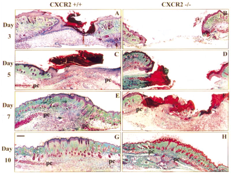
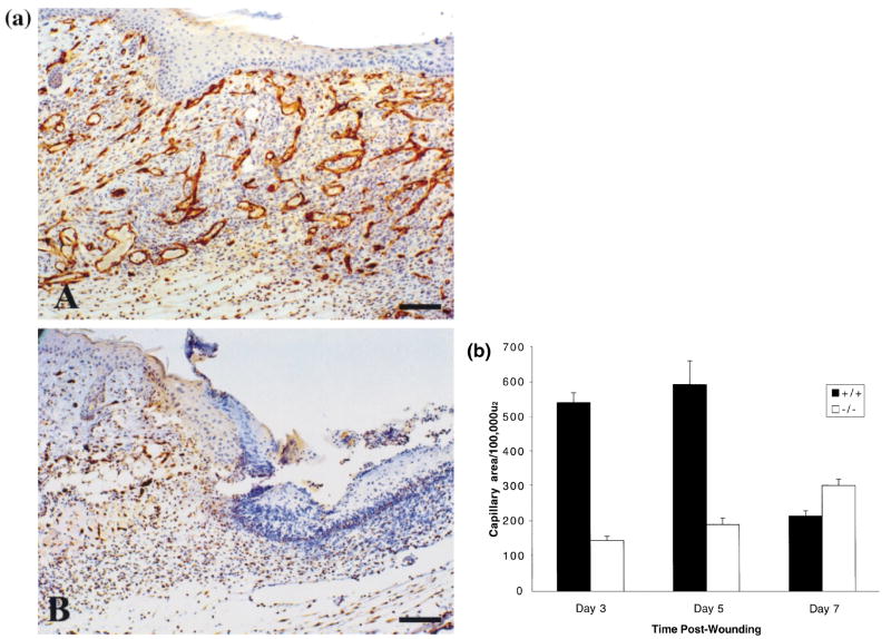
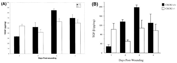
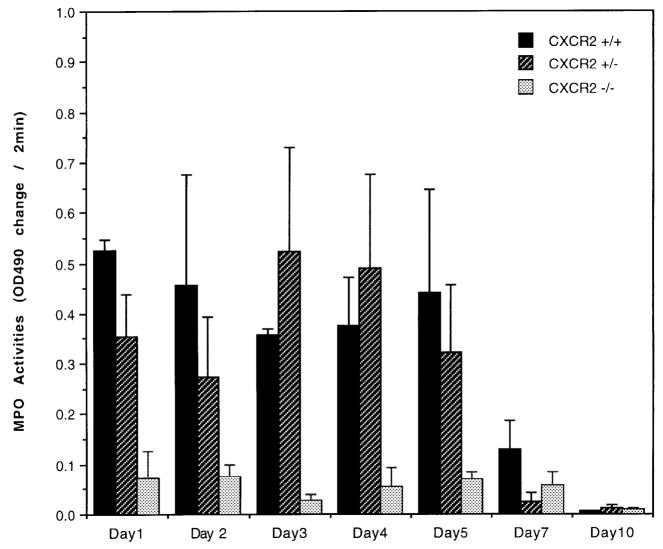
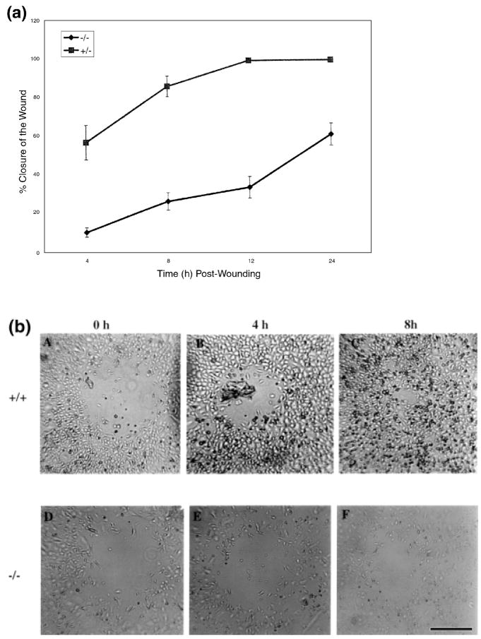
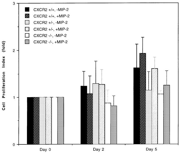
Previous studies demonstrated that the CXC chemokine, MGSA/GRO-alpha and its receptor, CXCR2, are expressed during wound healing by keratinocytes and endothelial cells at areas where epithelialization and neovascularization occur. The process of wound healing is dependent on leukocyte recruitment, keratinocyte proliferation and migration, and angiogenesis. These processes may be mediated in part by CXC chemokines, such as interleukin-8 and MGSA/GRO-alpha. To examine further the significance of CXC chemokines in wound healing, full excisional wounds were created on CXCR2 wild-type (+/+), heterozygous (+/-), or knockout (-/-) mice. Wounds were histologically analyzed for neutrophil and monocyte infiltration, neovascularization and epithelialization at days 3, 5, 7, and 10 postwounding. The CXCR2 -/- mice exhibited defective neutrophil recruitment, an altered temporal pattern of monocyte recruitment, and altered secretion of interleukin-1beta. Significant delays in wound healing parameters, including epithelialization and decreased neovascularization, were also observed in CXCR2 -/- mice. In vitro wounding experiments with cultures of keratinocytes established from -/- and +/+ mice revealed a retardation in wound closure in CXCR2 -/- keratinocytes, suggesting a role for this receptor on keratinocytes in epithelial resurfacing that is independent of neutrophil recruitment. These in vitro and in vivo studies further establish a pathophysiologic role for CXCR2 during cutaneous wound repair.


Figures

Figure 1
Figure 1. Delayed epithelial resurfacing and wound closure in CXCR2 −/− mice
Representative micrographs from trichrome-stained sections in parts (A)– (H) show responses to excisional injury on days 3, 5, 7, and 10 postinjury. Parts (A), (C), (E), and (G) depict the normal sequence of repair in +/+ (wild-type) mice. Epithelialization underneath the vivid red scab is nearly complete by day 3 with a hypertrophic epidermis by day 5. Robust cellularity in the wound bed, increasing collagen deposition, and wound contracture are evident at days 5, 7, and 10. By contrast, wounds from CXCR2 knockout mice shown in (B), (D), (F), and (H) reveal impaired reparative responses. Resurfacing across the wound bed remains incomplete until days 7–8. Severely inhibited cellularity of the granulation tissue (gt) is noted at days 3–5 and delays in wound contracture are evident at days 3, 5, and 7. Immature granulation tissue is visible at day 7 followed by eventual wound closure and complete resurfacing by day 10. All wounds eventually show complete epidermal and dermal healing with no scarring. After resurfacing is achieved, sites of excisional wounding can only be detected by a discontinuity in the underlying panniculus carnosus layer (pc). Scale bars: 100 μm.
Figure 2
Figure 2. (a) Immunostaining for CD31 antigen outlining blood vessels within 5 d wounds
(A) Numerous capillaries within the granulation tissue and 100% resurfacing by epithelium in CXCR2+/+ mice at postwound day 5. (B) Paucity of capillaries and incomplete resurfacing at the left and eschar at the top right. Scale bar. 100μm. (b) Delayed neovascularization in CXCR2−/− mice: Quantitative expression of capillary areas within the granulation tissue of excisional punches was assessed from hematoxylin and eosin sections as described in Materials and Methods. Values are expressed as the mean total capillary area per 100,000 (μm2 ± SEM at days 3, 5, and 7 postinjury, based upon five observations per point. CXCR2−/− mice exhibited significantly impaired neovascularization whereas CXCR2+/+ mice showed an expected peak in capillary in-growth followed by a regression in the capillary network that accompanies wound maturation (p < 0.0004, p < 0.003, p < 0.008 for postwound days 3, 5, and 7, respectively, Student’s two-tailed t test; or p = 2 E-09, p = 0.0002, and p = 0.05 for days 3, 5, and 7, respectively, by ANOVA).
Figure 3
Figure 3. Altered cytokine profiles in CXCR2−/− mice
VEGF (A) and TGF-β (B) levels in the wounded skin homogenates of CXCR2+/+ show a consistent rise and fall in these cytokines in wild-type mice from day 1 to day 4 postwounding. In CXCR2−/− mice the levels of TGF-β and VEGF were higher than for wild-type mice on postwound day 1. Values represent mean ± SEM of duplicate determinations for two to four mice per time point. Statistical differences in VEGF and TGF-β levels between wild-type and CXCR2−/− mice were noted on days 1 and 3, based upon the Student’s t test and ANOVA.
Figure 4
Figure 4. Reduced MPO activity in CXCR2−/− mice
Neutrophil MPO activity was measured on 50 μg protein from wound lysate. as described in Materials and Methods on tissue homogenates from postwound day 1 to day 10. Data are graphed as change in absorbance/optical density over a period of 2 min at 490 nm wavelength each day. The bars indicate the mean ± SEM from wounds of three to four wounded animals. In the CXCR2−/− mice MPO activity is significantly lower than in wild-type mice at postwound days 1, 3, and 5, based upon the Student’s two-tailed t test and/or ANOVA. MPO activity from wild-type mice is different from CXCR2 heterozygous mice (+/−) at postwound days 1 and 2 (p<0.05, Student’s two-tailed t test).
Figure 5
Figure 5. Delayed in vitro wound closure in keratinocytes from CXCR2−/− mice
In vitro wounding of confluent cultures of CXCR2+/−, and −/− keratinocytes. Keratinocytes from the three genotypes (+/+, +/−, and −/−) were isolated and cultured in 24 well tissue culture plates as described in Materials and Methods. Wounds (400–500 μm) on monolayered keratinocytes were monitored over a period of 24 h and the area of the wound defect determined using Bioquant software. Keratinocytes from wild-type mice exhibited a wound closure which was not significantly different from keratinocytes of heterozygous mice. Part (a) shows the percentage of wound closure of keratinocytes from +/− mice compared with −/− mice during a 24 h period. Values represent mean ± SEM obtained from cultures derived from three different mice and five independent wounds in each well. Part (b) depicts representative in vitro photographs of wounded keratinocyte cultures from +/+ (A–C) and −/− (D–F) mice during wound closure. Scale bar. 200 μm.
Figure 6
Figure 6. Effect of MIP-2 on the growth of keratinocytes from CXCR2+/+, +/−, and −/− mice
Keratinocyte cultures were established from newborn CXCR2+/+, +/−, and −/− mice according to the protocol described in Materials and Methods. Cell proliferation in the presence or absence of MIP-2 (100 ng per ml) was followed over 5 d by hemocytometer counting. Each point represents the mean of four individual determinations. The cell proliferation index on days 2 and 5 is defined as the mean number of cells per well divided by the mean number of cells attached 24 h after the initial seeding. The day the initial number of attached cells was determined is the time for the first addition of MIP-2 and this day was defined as day 0. The error bars represent the SEM.

References

    1. Abbot RE, Corral CJ, Maclvor DM, Lin X, Ley TJ, Mustoe TA. Augmented inflammatory response and altered wound healing in cathepsin G-deficient mice. Arch Surg. 1998;133:1002–1006. - PubMed
    1. Addison CL, Daniel TO, Ehlert JE, et al. The CXC. chemokine receptor 2, CXCR2, is the putative receptor for ELR+ CXC chemokine induced angiogenic activity. J Immunol. accepted. - PubMed
    1. Brauchle M, Fassler R, Werner S. Suppression of keratinocyte growth factor expression by glucocorticoids in vitro and during wound healing. J Invest Dermatol. 1995;105:579–584. - PubMed
    1. Breuss JM, Gallo J, DeLisser HM, et al. Expression of the beta 6 integrin subunit in development, neoplasia and tissue repair suggests a role in epithelial remodeling. J Cell Sci. 1995;108:2241–2251. - PubMed
    1. Brown LF, Yeo K, Berse B, Yeo T, Senger DR, Dvorak HF, Water LV. Expression of vascular permeability factor (Vascular Endothelial Growth Factor) by epidermal keratinocytes during wound healing. J Exp Med. 1992;176:1375–1379. - PMC - PubMed

Publication types

MeSH terms