Comparative Study
. 2000 Aug;130(8):1825-32.
doi: 10.1038/sj.bjp.0703506.

Ligand regulation of green fluorescent protein-tagged forms of the human beta(1)- and beta(2)-adrenoceptors; comparisons with the unmodified receptors

Affiliations
Comparative Study

Ligand regulation of green fluorescent protein-tagged forms of the human beta(1)- and beta(2)-adrenoceptors; comparisons with the unmodified receptors

A J McLean et al. Br J Pharmacol. 2000 Aug.

Abstract

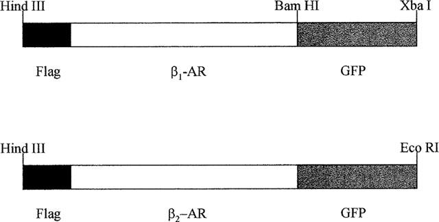
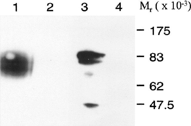
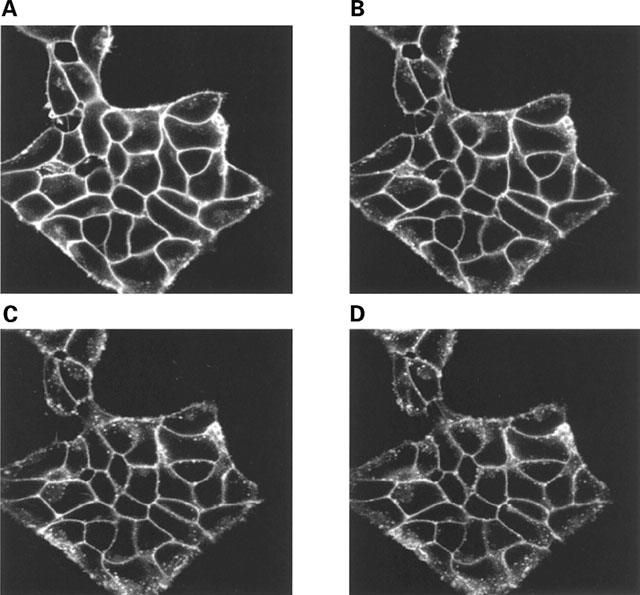
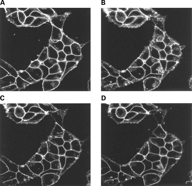
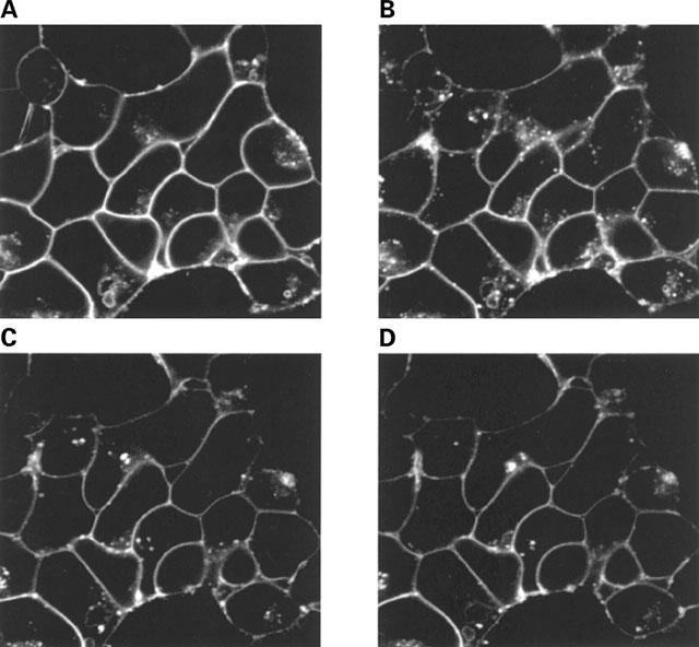
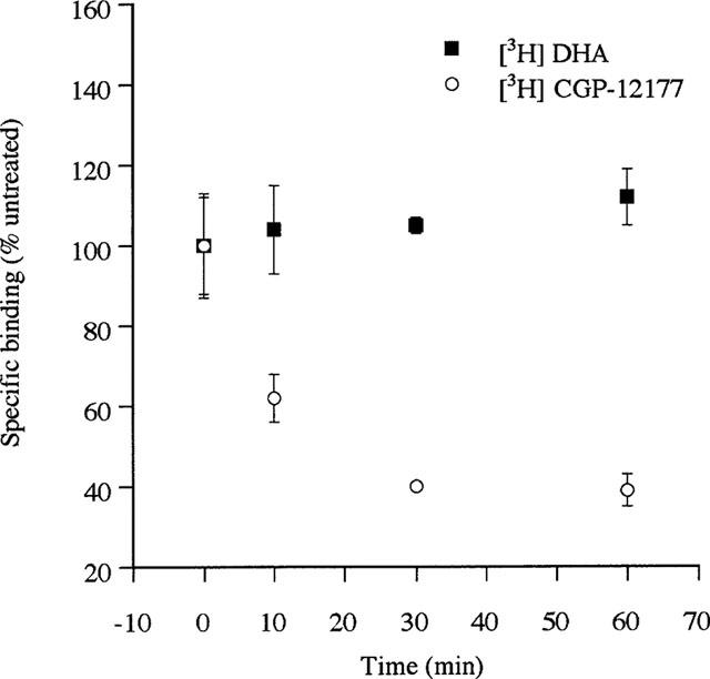
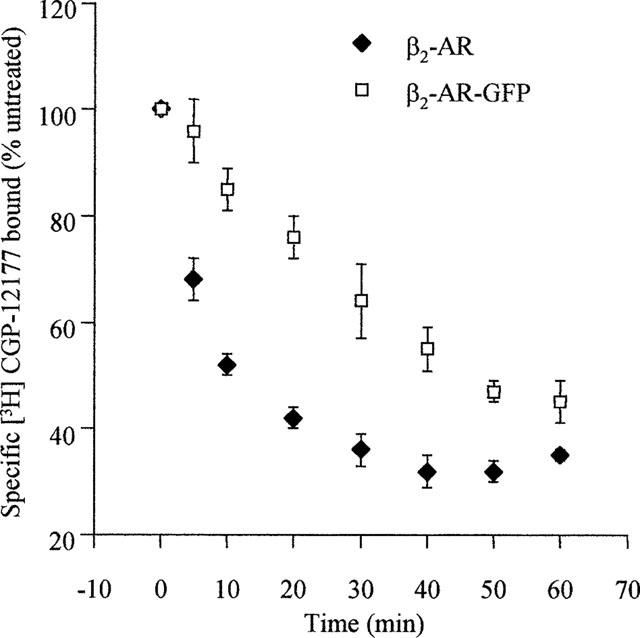
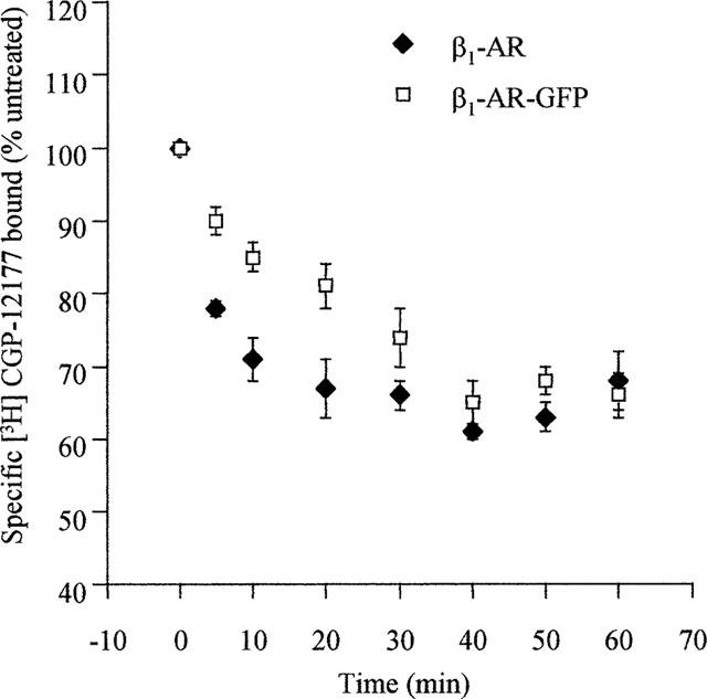
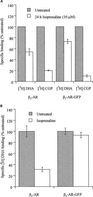
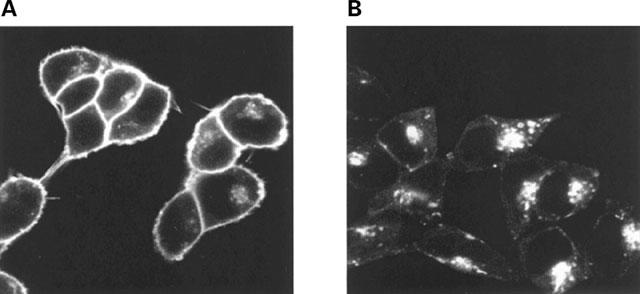
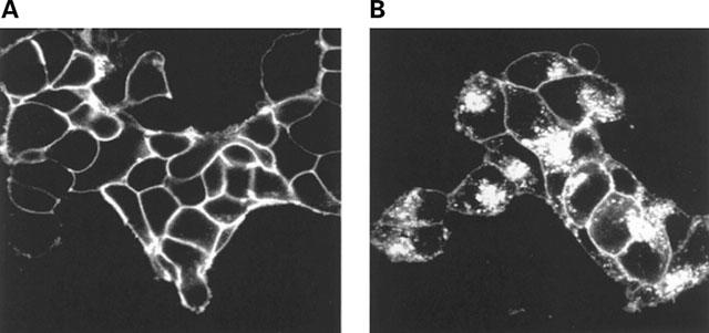
Stable clones of HEK293 cells expressing either FLAG(TM) epitope-tagged, wild type human beta(1)- and beta(2)-adrenoceptors or C-terminally green fluorescent protein (GFP)-tagged forms of these receptors were established. The binding affinity of [(3)H]-dihydroalprenolol and other ligands was little affected by addition of GFP to the C-terminal of either receptor. Isoprenaline induced the internalisation of both beta(1)-adrenoceptor-GFP and beta(2)-adrenoceptor-GFP and following removal of the agonist both constructs were able to recycle to the cell surface. The extent of internalisation of beta(2)-adrenoceptor-GFP produced by isoprenaline was substantially greater than for beta(1)-adrenoceptor-GFP. C-terminal addition of GFP slowed markedly the rate of internalization of both the beta(1)-adrenoceptor and the beta(2)-adrenoceptor in response to isoprenaline. Sustained exposure to isoprenaline (24 h) produced substantially greater levels of downregulation of native beta(2)-adrenoceptor compared to beta(2)-adrenoceptor-GFP although both were equally effectively removed from the plasma membrane. Sustained exposure to isoprenaline resulted in a large fraction of beta(2)-adrenoceptor-GFP becoming trapped in internal vesicles/lysosomes but not degraded. Even after sustained exposure to isoprenaline a significant fraction of beta(1)-adrenoceptor-GFP remained at the cell surface. These results indicate that although GFP tagging of beta-adrenoceptors can provide qualitative visual patterns of agonist-induced receptor trafficking and regulation in HEK293 cells the quantitative details vary markedly from those obtained with the unmodified receptors.


Figures

Figure 1
Figure 1
Schematic diagram of the constructs which were stably expressed in HEK293 cells. cDNAs encoding the human β1- and β2-adrenoceptor (AR) were modified at their N-terminus to encode a FLAGTM epitope. Following removal of the stop codon from the receptor sequences GFP was added in-frame.
Figure 2
Figure 2
Immunoblots of β1-adrenoceptor and β2-adrenoceptor constructs. Membranes expressing FLAGTM β2-adrenoceptor-GFP (1 and 4) or FLAGTM β1-adrenoceptor-GFP (2 and 3) were immunoblotted using either anti-β2-adrenoceptor (1 and 2) or anti-β1-adrenoceptor (3 and 4) antisera. Similar results were obtained in another set of immunoblots.
Figure 3
Figure 3
The β1-adrenoceptor-GFP construct is internalized following addition of isoprenaline. A group of cells stably expressing the FLAGTM β1-adrenoceptor-GFP construct were exposed to isoprenaline (10 μM) for 0 (A), 10 (B), 20 (C) or 40 (D) min during visualization in the confocal microscope. Three separate experiments produced similar results.
Figure 4
Figure 4
Replacement of isoprenaline with betaxolol allows the β1-adrenoceptor-GFP construct to recycle to the plasma membrane. Cells stably expressing the FLAGTM β1-adrenoceptor-GFP construct were exposed to isoprenaline (10 μM) for 0 (A) or 30 (B–D) min. Following washing in the presence of betaxolol (10 μM) the distribution of the receptor was visualized 10 (C) and 30 (D) min later. Three separate experiments produced similar results.
Figure 5
Figure 5
Internalization and recycling of β2-adrenoceptor-GFP. Cells stably expressing the FLAGTM β2-adrenoceptor-GFP construct were exposed to isoprenaline (10 μM) for 0 (A) or 30 (B–D) min. Following washing with buffer containing alprenolol (10 μM) the distribution of the receptor was monitored 30 (C) and 40 (D) min later. Three separate experiments produced similar results.
Figure 6
Figure 6
Short-term treatment with isoprenaline internalizes but does not downregulate the FLAGTM β2-adrenoceptor-GFP construct. Cells stably expressing the FLAGTM β2-adrenoceptor-GFP construct were exposed to isoprenaline (10 μM) for varying times. Specific binding of a single concentration of either [3H]-DHA (2 nM) or [3H]-CGP-12177 (10 nM) to intact cells was then measured as described in Methods.
Figure 7
Figure 7
Comparison of the kinetics and extent of isoprenaline-induced internalization of the β2-adrenoceptor and β2-adrenoceptor-GFP. Cells stably expressing either the FLAGTM β2-adrenoceptor or the FLAGTM β2-adrenoceptor-GFP construct were exposed to isoprenaline (10 μM) for varying times. Specific binding of a single concentration of [3H]-CGP-12177 (10 nM) to intact cells was then measured as described in Methods. Results are presented as means±s.e.m. for five (FLAGTM β2-adrenoceptor) or four (FLAGTM β2-adrenoceptor-GFP) experiments.
Figure 8
Figure 8
Comparison of the kinetics and extent of isoprenaline-induced internalization of the β1-adrenoceptor and β1-adrenoceptor-GFP. Cells stably expressing either the FLAGTM β1-adrenoceptor or the FLAGTM β1-adrenoceptor-GFP construct were exposed to isoprenaline (10 μM) for varying times. Specific binding of a single concentration of [3H]-CGP-12177 (10 nM) to intact cells was then measured as described in Methods. Results are presented as means±s.e.m. for three (FLAGTM β1-adrenoceptor) or five (FLAGTM β1-adrenoceptor-GFP) experiments.
Figure 9
Figure 9
Long-term treatment with isoprenaline. Effects on distribution and downregulation of the β2-adrenoceptor and β2-adrenoceptor-GFP monitored by ligand binding studies. (A) Intact Cells. Cells stably expressing either the FLAGTM β2-adrenoceptor or the FLAGTM β2-adrenoceptor-GFP construct were either untreated or exposed to isoprenaline (10 μM) for 24 h. Specific binding of a single concentration of either [3H]-DHA (2 nM) or [3H]-CGP-12177 (10 nM) to intact cells was then measured as described in Methods. Results are presented as means±s.e.m. for three separate experiments. (B) Membranes. Cells stably expressing either the FLAGTM β2-adrenoceptor or the FLAGTM β2-adrenoceptor-GFP construct were either untreated or exposed to isoprenaline (10 μM) for 24 h. Membranes were prepared and the specific binding of a single concentration of [3H]-DHA (2 nM) was then measured as described in Methods. Results are presented as means±s.e.m. for three separate experiments.
Figure 10
Figure 10
Visualization of the location of β2-adrenoceptor-GFP following long-term challenge with isoprenaline. Cells stably expressing the FLAGTM β2-adrenoceptor-GFP construct were either untreated (A) exposed to isoprenaline (10 μM) for 24 h (B). The distribution of the construct was then visualized. Two further experiments produced similar patterns.
Figure 11
Figure 11
Visualization of the location of β1-adrenoceptor-GFP following long-term challenge with isoprenaline. Cells stably expressing the FLAGTM β1-adrenoceptor-GFP construct were either untreated (A) exposed to isoprenaline (10 μM) for 24 h (B) The distribution of the construct was then visualized. Two further experiments produced similar patterns.

References

    1. AWAJI T., HIRASAWA H., KATAOKA M., SHINOURA H., NAKAYAMA Y., SUGAWARA T., IZUMI S., TSUJIMOTO G. Real-time optical monitoring of ligand-mediated internalisation of α1b-adrenoceptor with green fluorescent protein. Mol. Endocrinol. 1998;12:1099–1111. - PubMed
    1. BARAK L.S., FERGUSON S.S., ZHANG J., MARTENSON C., MEYER T., CARON M.G. Internal trafficking and surface mobility of a functionally intact beta2-adrenergic receptor-green fluorescent protein conjugate. Mol. Pharmacol. 1997;51:177–184. - PubMed
    1. BARLIC J., KHANDAKER M.H., MAHON E., ANDREWS J., DEVRIES M.E., MITCHELL G.B., RAHIMPOUR R., TAN C.M., FERGUSON S.S.G., KELVIN D.J. β-arrestins regulate interleukin-8-induced CXCR1 internalization. J. Biol. Chem. 1999;274:16287–16294. - PubMed
    1. CAO T.T., DEACON H.W., RECZEK D., BRETSCHER A., VON ZASTROW M. A kinase-regulated PDZ-domain interaction controls endocytic sorting of the β2-adrenergic receptor. Nature. 1999;401:286–290. - PubMed
    1. CONWAY B.R., MINOR L.K., XU J.Z., GUNNET J.W., DEBIASIO R., D'ANDREA M.R., RUBIN R., DEBIASIO R., GIULIANO K., ZHOU L., DEMAREST K.T. Quantification of G-protein coupled receptor internalization using G-protein coupled receptor-green fluorescent protein conjugates with the Array ScanTM high-content screening system. J. Biomol. Screening. 1999;4:75–86. - PubMed

Publication types

MeSH terms