. 2000 Aug 21;192(4):495-506.
doi: 10.1084/jem.192.4.495.

Subsecond induction of alpha4 integrin clustering by immobilized chemokines stimulates leukocyte tethering and rolling on endothelial vascular cell adhesion molecule 1 under flow conditions

Affiliations

Subsecond induction of alpha4 integrin clustering by immobilized chemokines stimulates leukocyte tethering and rolling on endothelial vascular cell adhesion molecule 1 under flow conditions

V Grabovsky et al. J Exp Med. .

Abstract

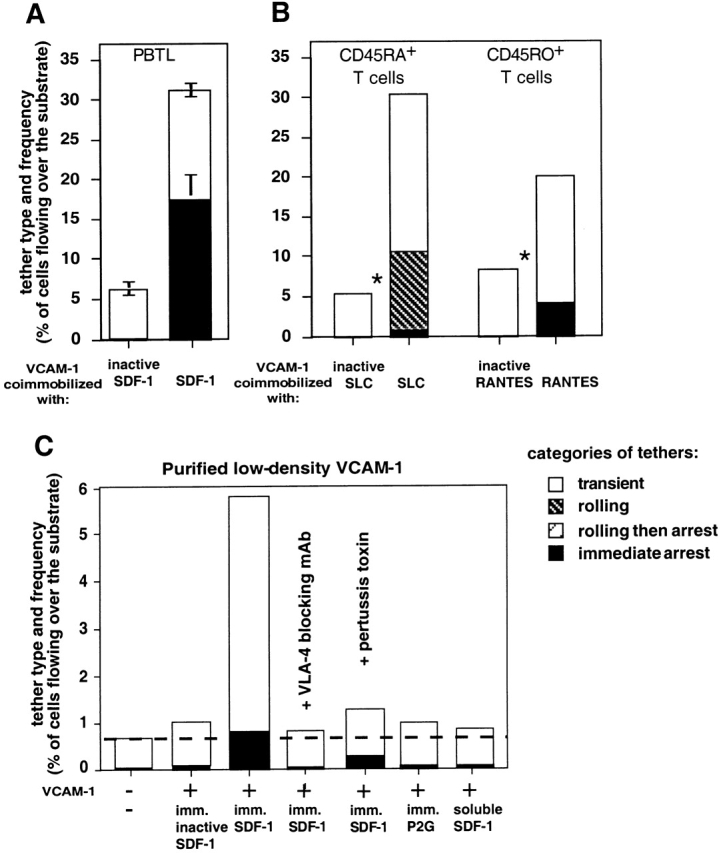
Leukocyte recruitment to target tissue is initiated by weak rolling attachments to vessel wall ligands followed by firm integrin-dependent arrest triggered by endothelial chemokines. We show here that immobilized chemokines can augment not only arrest but also earlier integrin-mediated capture (tethering) of lymphocytes on inflamed endothelium. Furthermore, when presented in juxtaposition to vascular cell adhesion molecule 1 (VCAM-1), the endothelial ligand for the integrin very late antigen 4 (VLA-4, alpha4beta1), chemokines rapidly augment reversible lymphocyte tethering and rolling adhesions on VCAM-1. Chemokines potentiate VLA-4 tethering within <0.1 s of contact through Gi protein signaling, the fastest inside-out integrin signaling events reported to date. Although VLA-4 affinity is not altered upon chemokine signaling, subsecond VLA-4 clustering at the leukocyte-substrate contact zone results in enhanced leukocyte avidity to VCAM-1. Endothelial chemokines thus regulate all steps in adhesive cascades that control leukocyte recruitment at specific vascular beds.


Figures

Figure 1
Figure 1
Cell surface–bound SDF-1 augments VLA-4–mediated capture and arrest of lymphocytes on endothelial VCAM-1 under physiological shear flow. (A) The frequency of PBTLs perfused at a shear stress of 1.5 dyn/cm2 over HUVECs stimulated for either 18 h (top) or 40 h (bottom) are capable of tethering transiently, or tether and roll or arrest on the cell monolayers. These different categories are depicted in stacked bars. SDF-1 (1 μg/ml in binding medium) was overlaid for 10 min on each monolayer and washed extensively before PBTL perfusion. Indicated cell monolayers were pretreated for 10 min with the E-selectin blocking mAb BB11 (at 10 μg/ml). Indicated PBTL samples were pretreated with the α4-integrin subunit mAb, HP1/2, to block their VLA-4–dependent interactions with endothelial VCAM-1. Where indicated, PBTLs were perfused over the endothelial monolayers in the presence of 1 mM EDTA. Standard deviation of total tethering values between multiple experiments on the different TNF-activated HUVECs was <10% of the mean. Asterisk indicates that chemokine-dependent augmentation in total tethering to activated HUVECs was highly significant (n = 5, P < 0.001). (B) Frequency of different categories of tethers initiated by PBTLs on VCAM-1–expressing CHO cells at 1.5 dyn/cm2. SDF-1 (1 μg/ml) was overlaid on the CHO monolayer as described for panel A. Data shown in B are representative of four independent experiments.
Figure 2
Figure 2
Immobilized chemokines augment VLA-4–mediated capture and arrest of T lymphocytes to purified VCAM-1 under shear flow. (A) Frequency of PBTL tethers to purified sVCAM-1 coated at 1.5 μg/ml on polystyrene surface with functional or heat-inactivated SDF-1 (2 μg/ml). The different tether categories were determined in two fields at 1 dyn/cm2 and results are an average and range of each tether category. (B) Frequency and type of tethers formed by naive (CD45RA+) and memory (CD45RO+) subsets of peripheral blood CD3+ T lymphocytes interacting at 1 dyn/cm2 with sVCAM-1 (1.5 μg/ml) coated together with the indicated intact or heat-inactivated chemokines (each at 2 μg/ml). Chemokine-dependent augmentation in total tethering was highly significant (*P < 0.001 and 0.004, for experiments performed on the CD45RA+ CD45RO+ subsets, respectively). (C) Frequency of VLA-4–mediated tethers of PBTLs to sVCAM-1 coated at 0.5 μg/ml, alone, or with inactivated SDF-1, SDF-1, or the non-signaling SDF-1 mutant P2G 16 (each at 2 μg/ml), determined at 0.5 dyn/cm2. VCAM-1 coating density on all substrates was identical as verified by radioimmunodetermination. For PTX treatment, PBTLs were cultured for 15 h with 100 ng/ml of the toxin. In A and D, where indicated, PBTLs were pretreated with the VLA-4 blocking mAb, HP1/2 (10 μg/ml), and were perfused unwashed over the VCAM-1–bearing surfaces. Data in A–C are representative of four independent experiments.
Figure 3
Figure 3
Effect of chemokines on VLA-4-mediated rolling of PBTLs on VCAM-1. (A) Lymphocytes were perfused at a shear stress of 1 dyn/cm2 on VCAM-1 (3 μg/ml) coimmobilized with inactivated or intact SDF-1 (1 μg/ml). The frequency of different categories of tethers is depicted in stacked bars. The contribution of high affinity VLA-4 subsets on perfused lymphocytes to each category was assessed by selective blocking of these subsets with EILDVPST peptide (at 0.75 mM). Data shown is representative from one of four experiments using different donors. (B) Chemokine triggering of VLA-4–dependent capture events followed by rolling or immediate arrests of PBTLs on VCAM-1, determined at 1.5 dyn/cm2 on VCAM-1 (3 μg/ml) coimmobilized with inactive SDF-1 or different intact chemokines (each at 2 μg/ml). (C) Instantaneous velocities of representative PBTLs interacting with VCAM-1 (3 μg/ml) coimmobilized with either inactive SDF-1 or active SDF-1 or IP-10 (1 and 2 μg/ml, respectively). The instantaneous velocities are plotted from the downstream entry of each cell into the field.
Figure 4
Figure 4
Immobilized chemokine and PMA modulate VLA-4 tether properties through distinct mechanisms. Kinetics of formation and dissociation of transient VLA-4–mediated tethers on very low density VCAM at a shear stress of 0.5 dyn/cm2. Effect of immobilized SDF-1 (A) or soluble PMA (B) on tether frequency and lifetime. (A) The duration of all tethers formed by equal number of Jurkat cells perfused for 1 min over sVCAM-1 (18 sites/μm2) alone or cocoated with SDF-1 at 2 μg/ml was determined, and the natural log of the tethers that remained bound after initiation of tethering was plotted against tether duration. (B) Effect of PMA treatment of Jurkat on the frequency and duration of Jurkat tethers to sVCAM-1 (36 sites/μm2). In A and B a first order dissociation fitting of tether duration is indicated by white symbols. Filled symbols denote longer tethers with high order dissociation kinetics. (C) Effect of immobilized SDF-1 and VCAM-1 density on frequency and duration of tethers formed by PBTLs interacting at a shear stress of 0.5 dyn/cm2 with the indicated densities of sVCAM-1 coimmobilized together with either inactivated or intact SDF-1 at 2 μg/ml. Least square analysis values of linear plots are depicted in r2 in A–C. Background tethering to HSA-coated substrate was 0.5% in A–C. Results are representative of three to four independent experiments.
Figure 5
Figure 5
Immobilized chemokine induces rapid clustering of VLA-4 at adhesive contact zones. (A) Effect of VCAM-1 density on frequency of PBTL tethering, measured at a shear stress of 0.5 dyn/cm2. All tethers measured on the indicated VCAM-1 densities were transient and diminished below a threshold VCAM-1 density (180 sites/μm2). (B) Confocal microscopy analysis of immunofluorescence staining of VLA-4 on PBTLs briefly incubated with control (HSA-coated) or SDF-1–coated beads (control and SDF-1, respectively), washed, fixed, and stained with the nonblocking VLA-4–specific mAb, B5G10. For PMA stimulation, cells were incubated with PMA for 2 min before VLA-4 staining. (C) Microclustering of VLA-4 and CXCR4 is enhanced within subsecond contact of PBTLs with surface-bound mAbs in the presence of immobilized SDF-1 leading to increased lymphocyte tethering to the surface. Tethering frequency of PBTLs measured at 0.75 dyn/cm2 to the VLA-4–, CXCR4-, or L-selectin–specific mAbs (HP1/2, 12G5, and DREG200, respectively), coated onto the substrates at 0.2 μg/ml, together with inactive SDF-1 (−), intact SDF-1, or the P2G SDF-1 mutant (+), or the control chemokine ELC, each at 2 μg/ml. The frequency of transient tethers and of tethers resulting in immediate arrests is depicted. The majority of transient tethers lasted <1 s. The non-PBTL binding mAb 4B9 (anti–VCAM-1) served as negative control. SDF-1–dependent augmentation in total tethering to anti–VLA-4 mAb coimmobilized with intact SDF-1 was significant compared with tethering measured in the presence of P2G (n = 4, P < 0.01). PTX pretreatment of PBTLs abolished 90 ± 5% of SDF-1–triggered PBTL tethering to the anti–VLA-4 mAb HP1/2 coimmobilized with intact SDF-1. (D) Effect of inhibitors to major integrin or GPCR signaling effectors on SDF-1–triggered tethering to VCAM-1. PBTLs were preincubated for 30 min with the PTK inhibitor (genestein; 100 μM), the PI-3K inhibitor (wortmannin; 100 nM), or with control DMSO solution (0.1%). VLA-4–dependent tethers were determined at 1 dyn/cm2 on VCAM-1 (1.5 μg/ml) coimmobilized with inactive or active SDF-1 (2 μg/ml, left). The effect of Ca2+ chelation on chemokine augmentation of VLA-4 tethering was tested on VCAM-1 (2 μg/ml) coimmobilized with SDF-1 (2 μg/ml) at shear stress of 1.5 dyn/cm2 (right). To chelate [Ca2+], PBTLs were preloaded with BAPTA-AM (at 25 μM) or control DMSO solution as described in Materials and Methods. The experiments shown in A–D are each representative of four independent experiments.
Figure 5
Figure 5
Immobilized chemokine induces rapid clustering of VLA-4 at adhesive contact zones. (A) Effect of VCAM-1 density on frequency of PBTL tethering, measured at a shear stress of 0.5 dyn/cm2. All tethers measured on the indicated VCAM-1 densities were transient and diminished below a threshold VCAM-1 density (180 sites/μm2). (B) Confocal microscopy analysis of immunofluorescence staining of VLA-4 on PBTLs briefly incubated with control (HSA-coated) or SDF-1–coated beads (control and SDF-1, respectively), washed, fixed, and stained with the nonblocking VLA-4–specific mAb, B5G10. For PMA stimulation, cells were incubated with PMA for 2 min before VLA-4 staining. (C) Microclustering of VLA-4 and CXCR4 is enhanced within subsecond contact of PBTLs with surface-bound mAbs in the presence of immobilized SDF-1 leading to increased lymphocyte tethering to the surface. Tethering frequency of PBTLs measured at 0.75 dyn/cm2 to the VLA-4–, CXCR4-, or L-selectin–specific mAbs (HP1/2, 12G5, and DREG200, respectively), coated onto the substrates at 0.2 μg/ml, together with inactive SDF-1 (−), intact SDF-1, or the P2G SDF-1 mutant (+), or the control chemokine ELC, each at 2 μg/ml. The frequency of transient tethers and of tethers resulting in immediate arrests is depicted. The majority of transient tethers lasted <1 s. The non-PBTL binding mAb 4B9 (anti–VCAM-1) served as negative control. SDF-1–dependent augmentation in total tethering to anti–VLA-4 mAb coimmobilized with intact SDF-1 was significant compared with tethering measured in the presence of P2G (n = 4, P < 0.01). PTX pretreatment of PBTLs abolished 90 ± 5% of SDF-1–triggered PBTL tethering to the anti–VLA-4 mAb HP1/2 coimmobilized with intact SDF-1. (D) Effect of inhibitors to major integrin or GPCR signaling effectors on SDF-1–triggered tethering to VCAM-1. PBTLs were preincubated for 30 min with the PTK inhibitor (genestein; 100 μM), the PI-3K inhibitor (wortmannin; 100 nM), or with control DMSO solution (0.1%). VLA-4–dependent tethers were determined at 1 dyn/cm2 on VCAM-1 (1.5 μg/ml) coimmobilized with inactive or active SDF-1 (2 μg/ml, left). The effect of Ca2+ chelation on chemokine augmentation of VLA-4 tethering was tested on VCAM-1 (2 μg/ml) coimmobilized with SDF-1 (2 μg/ml) at shear stress of 1.5 dyn/cm2 (right). To chelate [Ca2+], PBTLs were preloaded with BAPTA-AM (at 25 μM) or control DMSO solution as described in Materials and Methods. The experiments shown in A–D are each representative of four independent experiments.

References

    1. Springer T.A. Traffic signals for lymphocyte recirculation and leukocyte emigrationthe multistep paradigm. Cell. 1994;76:301–314. - PubMed
    1. Butcher E.C., Picker L.J. Lymphocyte homing and homeostasis. Science. 1996;272:60–66. - PubMed
    1. Kim C.H., Broxmeyer H.E. Chemokinessignal lamps for trafficking of T and B cells for development and effector function. J. Leukoc. Biol. 1999;65:6–15. - PubMed
    1. Gunn M.D., Tangemann K., Tam C., Cyster J.G., Rosen S.D., Williams L.T. A chemokine expressed in lymphoid high endothelial venules promotes the adhesion and chemotaxis of naive T lymphocytes. Proc. Natl. Acad. Sci. USA. 1998;95:258–263. - PMC - PubMed
    1. Campbell J.J., Haraldsen G., Pan J., Rottman J., Qin S., Ponath P., Andrew D.P., Warnke R., Ruffing N., Kassam N. The chemokine receptor CCR4 in vascular recognition by cutaneous but not intestinal memory T cells. Nature. 1999;400:776–780. - PubMed

Publication types

MeSH terms