. 2000 Aug 21;192(4):537-48.
doi: 10.1084/jem.192.4.537.

Premature expression of T cell receptor (TCR)alphabeta suppresses TCRgammadelta gene rearrangement but permits development of gammadelta lineage T cells

Affiliations

Premature expression of T cell receptor (TCR)alphabeta suppresses TCRgammadelta gene rearrangement but permits development of gammadelta lineage T cells

K Terrence et al. J Exp Med. .

Abstract

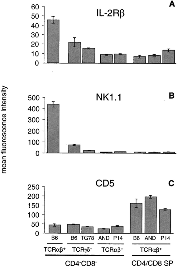
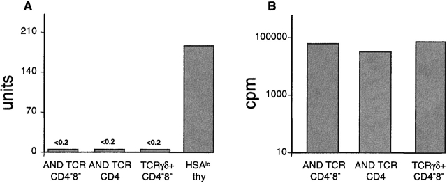
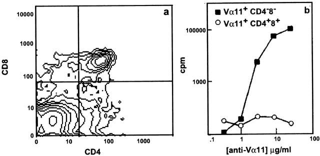
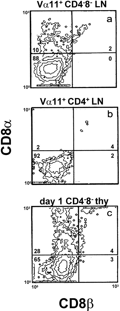
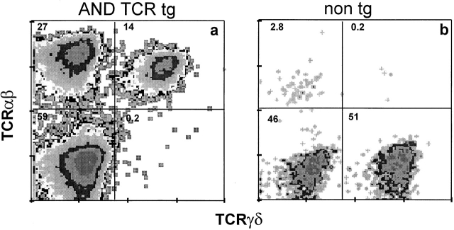
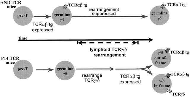
The T cell receptor (TCR)gammadelta and the pre-TCR promote survival and maturation of early thymocyte precursors. Whether these receptors also influence gammadelta versus alphabeta lineage determination is less clear. We show here that TCRgammadelta gene rearrangements are suppressed in TCRalphabeta transgenic mice when the TCRalphabeta is expressed early in T cell development. This situation offers the opportunity to examine the outcome of gammadelta versus alphabeta T lineage commitment when only the TCRalphabeta is expressed. We find that precursor thymocytes expressing TCRalphabeta not only mature in the alphabeta pathway as expected, but also as CD4(-)CD8(-) T cells with properties of gammadelta lineage cells. In TCRalphabeta transgenic mice, in which the transgenic receptor is expressed relatively late, TCRgammadelta rearrangements occur normally such that TCRalphabeta(+)CD4(-)CD8(-) cells co-express TCRgammadelta. The results support the notion that TCRalphabeta can substitute for TCRgammadelta to permit a gammadelta lineage choice and maturation in the gammadelta lineage. The findings could fit a model in which lineage commitment is determined before or independent of TCR gene rearrangement. However, these results could be compatible with a model in which distinct signals bias lineage choice and these signaling differences are not absolute or intrinsic to the specific TCR structure.


Figures

Figure 1
Figure 1
Phenotypic markers distinguish the TCRαβDN T cell subset of TCRαβ transgenic mice from αβ lineage T cells (CD4 and CD8), NK T, but not from γδ lineage T cells. Thymocytes (A and B) or B cell–depleted lymph node T cells (C) from B6, transgenic TCRγδ (TG78), or transgenic TCRαβ (AND and P14) mice were each stained for TCRαβ, TCRγδ, CD4 and/or CD8, and a fifth marker (IL-2Rβ, NK1.1, or CD5), and analyzed by flow cytometry. The mean fluorescence intensity for the specified markers was determined for CD4/CD8 single positive (SP) T cells by software gating for CD4+CD8TCRαβhi and CD4CD8+TCRαβhi, or for CD4CD8 T cells, by live gating for CD4CD8 followed by software gating for TCRαβ+ or TCRγδ+. Each bar represents the means (with SE bars) collected from analysis of three individual mice with the exception of B6 (two mice).
Figure 2
Figure 2
TCRαβDN T cells respond to anti-TCR stimulation by proliferating but do not produce an IL-4 response. Lymph node CD4CD8 or CD4+ T cells from AND TCR (Vα11/Vβ3), CD48TCRγδ+ T cells from G8 TCR mice (purified using magnetic beads and electronic cell sorting), or HSAlo B6 thymocytes (an enriched source of NK T cells), plated at 5 × 10⁴ cells/well, were stimulated with 10 μg/ml immobilized anti-TCR antibody (anti-Vα11 for AND TCR, anti-TCRγδ for G8 TCR, and anti-TCRαβ for B6 TCR) and assayed for (A) IL-4 production where 1 unit = 0.5 pg of IL-4, and for (B) proliferation. Data are representative of two experiments, averaging values from triplicate wells, and are derived from dose–response curves using 0.1–100 μg/ml of antibody.
Figure 3
Figure 3
TCRαβDN cells appear early in thymic development. Thymocytes were harvested day 15 after reconstituting B10.BR or B10.A RAG-2−/− irradiated recipients with T-depleted H-2k AND TCR bone marrow. (a) Cells were stained for CD4, CD8, and Vα11 TCR and analyzed by three-color flow cytometry. (b) Thymocytes were electronically sorted for Vα11+CD4+CD8+ and Vα11+CD4CD8. Sorted cells (12 × 104/well) were tested in a proliferation assay for response to plate-bound anti-Vα11 (RR8-1) antibody. Proliferation data are representative of two sorting experiments, and the cytometric analysis on day 15 is representative from several series of analyses performed on thymocytes from chimeric mice on days 10–20 after reconstitution.
Figure 4
Figure 4
TCR stimulation can induce CD8αα expression on TCRαβ DN T cells. (a) Vα11+ CD4CD8, (b) Vα11+CD4+ lymph node T cells from AND TCR mice (isolated by electronic cell sorting and cultured at 4 × 10⁴/well on 30 μg/ml plate-bound anti-Vα11, RR8-1, in the presence of recombinant IL-1 and IL-2, 100 U/ml, each), and (c) CD4CD8 thymocytes of day 1 neonatal mice (isolated by magnetic bead depletion and cultured at 10 × 104/well on 24 μg/ml immobilized anti-γδ, GL-3, in the presence of rIL-1 and rIL-2, 20 U/ml each) were assayed for expression of CD8α and CD8β by flow cytometry. The data are representative of three or more experiments. B6 LN T cells were used as a positive control for CD8β staining (data not shown).
Figure 5
Figure 5
TCRαβDN cells do not require MHC-dependent positive selection for development. Thymocytes were isolated from MHC class I–specific (HY and P14) or class II–specific (AND, 5CC7, and DO11.10) TCR transgenic mice bred onto a positively selecting (pos) or nonselecting, neutral (neut) MHC background. Cells were stained for CD4, CD8, and TCR (using antibodies: T370 for HY, anti-Vα2 for P14, anti-Vα11 for AND and 5CC7, and KJ-126 for DO11.10 TCR). The percent of transgenic (tg) TCRhi cells was determined from analysis of total thymocytes by software gating for CD4CD8, CD48+, or CD4+CD8. Each bar represents the mean percentage (with SE bars) of TCRhi of CD4CD8 or of total thymocytes from analyses of three to six individual mice per group.
Figure 6
Figure 6
CD4CD8 T cells of AND and HY TCR mice express transgenic TCRαβ but no TCRγδ, while those of P14 TCR mice coexpress transgenic TCRαβ and endogenous TCRγδ. Thymocytes and lymph node cells, stained for TCRαβ (H57-597), TCRγδ (GL3), CD4, and CD8, were analyzed by flow cytometry using live gating to collect data only from CD4CD8 cells. The numbers inside the quadrants represent the percentage of CD4CD8 thymocytes in each population. Statistics are given in Table .
Figure 7
Figure 7
The transgenic AND TCR is expressed much earlier than the P14 TCR in fetal development. Thymocytes from P14 TCR or AND TCR embryos, harvested on the days indicated (E13–18), were stained for Thy 1.2, CD4, CD8, and TCR (Vα2 for P14 and Vα11 for AND) and analyzed by four color flow cytometry. Distributions, gated for total Thy1.2+ thymocytes, display dual parameter, CD4 and CD8, or single parameter, TCR (shaded), overlaid with the negative control for background fluorescence (unshaded). Numbers indicate the percentage of cells within the indicated gates. Data are representative of two such experiments with similar time courses.
Figure 8
Figure 8
Vγ2–Jγ1 gene rearrangements are suppressed in TCR+ CD4CD8 (TCRαβDN) cells of AND TCR but not of P14 TCR mice. Rearrangements are suppressed, independent of MHC haplotype, in the TCRαβDN but not the TCR+CD4+CD8 subset of AND TCR mice. Samples of 105 each of TCR+ (Vα11+ for AND, Vα2+ for P14, TCRγδ+ for G8 TCR, and TCRγδ+ or TCRαβ+ for B6) (a) CD4CD8, (b) CD4+CD8, and (c) CD4CD8+ lymph node T cells from AND TCR/H-2b, AND TCR/H-2b (MHC class II+/−, CD4+/−), AND TCR/H-2q, P14 TCR, G8 TCR, and B6 mice were isolated by electronic sorting. The relative frequency of Vγ2–Jγ1 rearrangements per sample was determined using a PCR ELISA as described in Materials and Methods. Bars represent the mean values (with SEs) of three individual sorts, using a total of five to eight mice per sort.
Figure 9
Figure 9
In contrast to lymphoid T cells, skin dendritic epithelial lymphocytes of AND TCR mice contain two subsets of T cells, one bearing only the transgenic TCRαβ and a second coexpressing the transgenic TCRαβ with endogenous TCRγδ. Isolated epidermal lymphocytes of (a) H-2d AND TCR transgenic (tg) or (b) nontransgenic (non tg) B10.D2 mice were stained for TCRαβ (H57-597) and TCRγδ (GL3) and analyzed by flow cytometry. The numbers inside the quadrant represent the percentage of cells in each population. The data are representative of several analyses of AND TCR mice of H-2d or other H-2 haplotypes.
Figure 10
Figure 10
A model to explain the different patterns of TCR expression in CD4CD8 thymocytes of TCRαβ transgenic mice. Lymphoid-type TCRγδ gene rearrangements occur at a distinct time (stage) in thymocyte development. In AND TCR mice, the transgenic TCRαβ is expressed early with respect to TCRγδ gene rearrangement and rearrangement is suppressed. Since transgenic TCRαβ expression occurs somewhat later in P14 TCR mice, there is no interference with rearrangement and cells with in-frame TCRγδ rearrangements can express both the transgenic TCRαβ and endogenous TCRγδ. Irrespective of the TCR expression pattern, CD4CD8 cells mature with properties of γδ lineage T cells.

References

    1. Bluestone J.A., Cron R.Q., Barrett T.A., Houlden B., Sperling A.I., Dent A., Hedrick S., Rellahan B., Matis L.A. Repertoire development and ligand specificity of murine TCRγδ cells. Immunol. Rev. 1991;120:5–33. - PubMed
    1. Allison J. γδ T-cell development. Curr. Opin. Immunol. 1993;5:241–246. - PubMed
    1. von Boehmer H., Aifantis I., Azogui O., Feinberg J., Saint-Ruf C., Zober C., Garcia C., Buer J. Crucial function of the pre-T-cell receptor (TCR) in TCRβ selection, TCRβ allelic exclusion and αβ versus γδ lineage commitment. Immunol. Rev. 1998;165:111–119. - PubMed
    1. Kang J., Raulet D.H. Events that regulate differentiation of αβ TCR+ and γδ TCR+ T cells from a common precursor. Semin. Immunol. 1997;9:171–179. - PubMed
    1. Robey E., Fowlkes B.J. The αβ versus γδ T-cell lineage choice. Curr. Opin. Immunol. 1998;10:181–187. - PubMed

MeSH terms

Substances