. 2000 Aug 21;192(4):587-94.
doi: 10.1084/jem.192.4.587.

Macrophage-tropic HIV induces and exploits dendritic cell chemotaxis

Affiliations

Macrophage-tropic HIV induces and exploits dendritic cell chemotaxis

C L Lin et al. J Exp Med. .

Abstract

Immature dendritic cells (iDCs) express the CC chemokine receptor (CCR)5, which promotes chemotaxis toward the CC chemokines regulated on activation, normal T cell expressed and secreted (RANTES), macrophage inflammatory protein (MIP)-1alpha, and MIP-1beta. By contrast, mature DCs downregulate CCR5 but upregulate CXC chemokine receptor (CXCR)4, and as a result exhibit enhanced chemotaxis toward stromal cell-derived factor (SDF)-1alpha. CCR5 and CXCR4 also function as coreceptors for macrophage-tropic (M-tropic) and T cell-tropic (T-tropic) human immunodeficiency virus (HIV)-1, respectively. Here, we demonstrate chemotaxis of iDCs toward M-tropic (R5) but not T-tropic (X4) HIV-1. Furthermore, preexposure to M-tropic HIV-1 or its recombinant envelope protein prevents migration toward CCR5 ligands. The migration of iDCs toward M-tropic HIV-1 may enhance formation of DC-T cell syncytia, thus promoting viral production and destruction of both DC and T helper lymphocytes. Therefore, disturbance of DC chemotaxis by HIV-1 is likely to contribute to immunosuppression in primary infection and AIDS. In addition, migration of iDCs toward HIV-1 may aid the capture of R5 HIV-1 virions by the abundant DC cell surface protein DC-specific intercellular adhesion molecule (ICAM)3-grabbing nonintegrin (DC-SIGN). HIV-1 bound to DC cell-specific DC-SIGN retains the ability to infect replication-permissive T cells in trans for several days. Consequently, recruitment of DC by HIV-1 could combine with the ability of DC-SIGN to capture and transmit the virus to T cells, and so facilitate dissemination of virus within an infected individual.


Figures
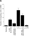
Figure 1
Figure 1
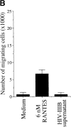
iDCs migrate toward RANTES and M-tropic HIV culture supernatant but not toward T-tropic HIV or uninfected supernatants. (A) Chemotaxis of 5 × 10⁴ immatures DCs in a bare transwell migration assay (described in Materials and Methods). The number of cells migrating into the lower transwells (×1,000) as determined by FACSort™ (Becton Dickinson) is shown with lower transwells containing medium alone (RPMI 1640), 6 nM RANTES, supernatant from PM1 T cells alone (which express CCR5), and PM1 cells infected with M-tropic HIV-1BaL. Cell supernatants were separated into concentrate (two times) and filtrate by centrifugation through a 100-kD cutoff centricon concentrator (Amicon). In all figures, the >100-kD fraction is used as “supernatant.” The >100-kD fraction from HIV-1BaL–infected PM1 cells is highly chemotactic, even when added in a 10-fold dilution; this large molecular mass chemotactic agent is likely HIV-virions/fragments and/or gp120 (see Fig. 3). (B) As in A, except using >100-kD supernatant from H9 T cells infected with the T-tropic HIV-1IIIB. In both graphs, SD from the mean is shown for four experiments, each with two transwells. Results are representative of four experiments
Figure 1
Figure 1
iDCs migrate toward RANTES and M-tropic HIV culture supernatant but not toward T-tropic HIV or uninfected supernatants. (A) Chemotaxis of 5 × 10⁴ immatures DCs in a bare transwell migration assay (described in Materials and Methods). The number of cells migrating into the lower transwells (×1,000) as determined by FACSort™ (Becton Dickinson) is shown with lower transwells containing medium alone (RPMI 1640), 6 nM RANTES, supernatant from PM1 T cells alone (which express CCR5), and PM1 cells infected with M-tropic HIV-1BaL. Cell supernatants were separated into concentrate (two times) and filtrate by centrifugation through a 100-kD cutoff centricon concentrator (Amicon). In all figures, the >100-kD fraction is used as “supernatant.” The >100-kD fraction from HIV-1BaL–infected PM1 cells is highly chemotactic, even when added in a 10-fold dilution; this large molecular mass chemotactic agent is likely HIV-virions/fragments and/or gp120 (see Fig. 3). (B) As in A, except using >100-kD supernatant from H9 T cells infected with the T-tropic HIV-1IIIB. In both graphs, SD from the mean is shown for four experiments, each with two transwells. Results are representative of four experiments
Figure 3
Figure 3
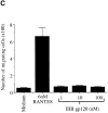
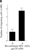
iDCs migrate toward recombinant M-tropic but not T-tropic gp120. (A) Chemotaxis of iDCs as described in the legend to Fig. 1 toward baculovirus expressed recombinant M-tropic gp120 from HIV-1BaL (n = 3). Migration toward HIV-1BaL gp120 was observed in 12 experiments. (B) Chemotaxis of iDCs as described in the legend to Fig. 1 toward baculovirus-expressed recombinant M-tropic gp120 from HIV-1ADA (n = 3). Data is representative of four experiments. (C) Chemotaxis of iDCs as described in the legend to Fig. 1 toward baculovirus-expressed recombinant T-tropic gp120 from HIV-1IIIB gp120 (available from the AIDS Reagent Project, Medical Research Council, UK). Data is representative of three experiments. Binding of gp120s to CD4 was confirmed by surface plasmon resonance (BIAcore). All graphs show SD from the mean for three transwells. Similar results were observed for gp120 from the T-tropic strains SF2, MN, W61D, and HXB2. (D) Addition of 1 μg of anti-CCR5 mAb 2D7 (BD PharMingen) or 45531.111 (R&D Systems) to the transwell insert inhibits migration to recombinant HIV-1BaL gp120. Graph shows the SD from the mean for three replicate transwells. Results are representative of four experiments.
Figure 3
Figure 3
iDCs migrate toward recombinant M-tropic but not T-tropic gp120. (A) Chemotaxis of iDCs as described in the legend to Fig. 1 toward baculovirus expressed recombinant M-tropic gp120 from HIV-1BaL (n = 3). Migration toward HIV-1BaL gp120 was observed in 12 experiments. (B) Chemotaxis of iDCs as described in the legend to Fig. 1 toward baculovirus-expressed recombinant M-tropic gp120 from HIV-1ADA (n = 3). Data is representative of four experiments. (C) Chemotaxis of iDCs as described in the legend to Fig. 1 toward baculovirus-expressed recombinant T-tropic gp120 from HIV-1IIIB gp120 (available from the AIDS Reagent Project, Medical Research Council, UK). Data is representative of three experiments. Binding of gp120s to CD4 was confirmed by surface plasmon resonance (BIAcore). All graphs show SD from the mean for three transwells. Similar results were observed for gp120 from the T-tropic strains SF2, MN, W61D, and HXB2. (D) Addition of 1 μg of anti-CCR5 mAb 2D7 (BD PharMingen) or 45531.111 (R&D Systems) to the transwell insert inhibits migration to recombinant HIV-1BaL gp120. Graph shows the SD from the mean for three replicate transwells. Results are representative of four experiments.
Figure 2
Figure 2
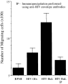
HIV envelope antibodies reduce the chemotaxis induced by M-tropic HIV. Immunoprecipitation (IP) of HIV-1BaL supernatant reduces the induced chemotaxis to the level of HIV-1IIIB supernatant. Similar immunoprecipitation of MIP-1β in RPMI resulted in only a 14% decrease in overall chemotaxis, controlling for general negative effects of either the antibodies or protein A sepharose. SEM is shown for three transwells.
Figure 4
Figure 4
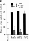
Exposure of iDCs to M-tropic HIV or its recombinant envelope protein inhibits subsequent chemotaxis toward CCR5 ligands but not toward the CXCR4 ligand SDF-1α. (A) iDCs were incubated for 90 min at 37°C in RPMI medium alone, or in supernatant from uninfected PM1 cells or supernatant from PM1 cells infected with HIV-1BaL before use in bare transwell assays, as described in the legend to Fig. 1. Graph shows SD from the mean for six transwells each. (B) Addition of 3 nM M-tropic HIVBaL gp120 (described in the legend to Fig. 3 A) to the transwell insert during a transwell assay (as described in the legend to Fig. 1) inhibits chemotaxis of iDCs toward the CCR5 ligand MIP-1β. SD from the mean is shown for three transwells. Data is representative of three separate experiments.
Figure 4
Figure 4
Exposure of iDCs to M-tropic HIV or its recombinant envelope protein inhibits subsequent chemotaxis toward CCR5 ligands but not toward the CXCR4 ligand SDF-1α. (A) iDCs were incubated for 90 min at 37°C in RPMI medium alone, or in supernatant from uninfected PM1 cells or supernatant from PM1 cells infected with HIV-1BaL before use in bare transwell assays, as described in the legend to Fig. 1. Graph shows SD from the mean for six transwells each. (B) Addition of 3 nM M-tropic HIVBaL gp120 (described in the legend to Fig. 3 A) to the transwell insert during a transwell assay (as described in the legend to Fig. 1) inhibits chemotaxis of iDCs toward the CCR5 ligand MIP-1β. SD from the mean is shown for three transwells. Data is representative of three separate experiments.

References

    1. Hussain L.A., Lehner T. Comparative investigation of Langerhans' cells and potential receptors for HIV in oral, genitourinary and rectal epithelia. Immunology. 1995;85:475–484. - PMC - PubMed
    1. Spira A.I., Marx P.A., Patterson B.K., Mahoney J., Koup R.A., Wolinsky S.M., Ho D.D. Cellular targets of infection and route of viral dissemination after an intravaginal inoculation of simian immunodeficiency virus into rhesus macaques. J. Exp. Med. 1996;183:215–225. - PMC - PubMed
    1. Knight S.C., Patterson S. Bone marrow-derived dendritic cells, infection with human immunodeficiency virus, and immunopathology. Annu. Rev. Immunol. 1997;15:593–615. - PubMed
    1. Inaba K., Steinman R.M. Accessory cell–T lymphocyte interactions. Antigen-dependent and -independent clustering. J. Exp. Med. 1986;163:247–261. - PMC - PubMed
    1. Cameron P.U., Freudenthal P.S., Barker J.M., Gezelter S., Inaba K., Steinman R.M. Dendritic cells exposed to human immunodeficiency virus type-1 transmit a vigorous cytopathic infection to CD4+ T cells. Science. 1992;257:383–387. - PubMed

Publication types

MeSH terms