. 2000 Aug 21;150(4):807-22.
doi: 10.1083/jcb.150.4.807.

Stimulation of fascin spikes by thrombospondin-1 is mediated by the GTPases Rac and Cdc42

Affiliations

Stimulation of fascin spikes by thrombospondin-1 is mediated by the GTPases Rac and Cdc42

J C Adams et al. J Cell Biol. .

Abstract

Cell adhesion to extracellular matrix is an important physiological stimulus for organization of the actin-based cytoskeleton. Adhesion to the matrix glycoprotein thrombospondin-1 (TSP-1) triggers the sustained formation of F-actin microspikes that contain the actin-bundling protein fascin. These structures are also implicated in cell migration, which may be an important function of TSP-1 in tissue remodelling and wound repair. To further understand the function of fascin microspikes, we examined whether their assembly is regulated by Rho family GTPases. We report that expression of constitutively active mutants of Rac or Cdc42 triggered localization of fascin to lamellipodia, filopodia, and cell edges in fibroblasts or myoblasts. Biochemical assays demonstrated prolonged activation of Rac and Cdc42 in C2C12 cells adherent to TSP-1 and activation of the downstream kinase p21-activated kinase (PAK). Expression of dominant-negative Rac or Cdc42 in C2C12 myoblasts blocked spreading and formation of fascin spikes on TSP-1. Spreading and spike assembly were also blocked by pharmacological inhibition of F-actin turnover. Shear-loading of monospecific anti-fascin immunoglobulins, which block the binding of fascin to actin into cytoplasm, strongly inhibited spreading, actin cytoskeletal organization and migration on TSP-1 and also affected the motility of cells on fibronectin. We conclude that fascin is a critical component downstream of Rac and Cdc42 that is needed for actin cytoskeletal organization and cell migration responses to thrombospondin-1.


Figures
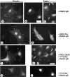
Figure 1
Figure 1
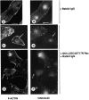
Effects of constitutively active Rho or Cdc42 on the localization of vinculin and fascin in quiescent fibroblasts. NIH 3T3 fibroblasts were stained with mouse monoclonal antibodies to vinculin (A, D, and F) or fascin (C, E, G, and H) 6 h after microinjection with rabbit IgG (A–C); IgG plus Q63L Rho plasmid (D and E); IgG plus Q61L Cdc42 and T17N Rac plasmids (F and G), or IgG plus T17N Rac plasmid alone (H and J). TRITC-anti–rabbit IgG staining was used to identify injected cells (exemplified by fields shown in A and B, H and J). Arrows in C–G indicate injected cells. Thin arrow in G indicates band of fascin at cell edge. Bars, 10 μm.
Figure 2
Figure 2
Effects of constitutively active Rac on the localization of vinculin and fascin in quiescent fibroblasts. NIH 3T3 fibroblasts were stained with mouse monoclonal antibodies to vinculin (A) or fascin (B and C) 6 h after injection with rabbit IgG plus Q61L Rac plasmid. The injected cells are indicated by arrows in each panel. Cells a and b in C illustrate different densities of the radial fascin staining intensities, cell c shows a cluster of short fascin projections and cell d illustrates the minority of cells which showed short projections rather than fascin ribbons as in B. Cell e in C is an injected cell not in the plane of focus. See text for further details. Bars: (A and B); 10 μm, (C) 5 μm.
Figure 3
Figure 3
Spatial relationship of Q61L Cdc42-stimulated actin projections and focal contacts in fibroblasts. NIH 3T3 fibroblasts were injected with IgG (A and B) or IgG plus Q61L Cdc42 and T17N Rac plasmids (C–F) and double-stained 6 h later for F-actin (A, C, and E) and vinculin (B, D, and F). The large projection arrowed in C and D shows vinculin at the tip. Examples of actin projections which do not contain organized focal contacts are indicated by arrows in E and F. Bars, 10 μm.
Figure 4
Figure 4
Distribution of fascin and phosphotyrosine-containing proteins in Cdc42 and Rac-stimulated cytoskeletal structures. C2C12 cells transiently cotransfected with Q61L Cdc42 and EGFP-fascin (A, B, E, F, and G) or Q61L Rac and EGFP-fascin (C and D) were plated on fibronectin for 60 min (A and B) or 30 min (C–G) and counterstained for F-actin (A) or phosphotyrosine (C, E, and G). Arrows in E and G indicate transfected cells. Bars: (A and B) 5 μm; (C–G) 10 μm.
Figure 5
Figure 5
Activation of Rac and Cdc42 by matrix adhesion. (A) Specificity of PDB binding for GTP-Rac and GTP-Cdc42. (B) The activity status of Cdc42 and Rac was examined by PBD binding assay in Triton X-100 or NP-40 extracts, respectively, of C2C12 myoblastic cells after adhesion to TSP-1 (T) or FN (F) for the indicated times, or after suspension for 90 min in BSA-blocked dishes (S). (C) Rac or Cdc42 protein levels were determined by Western blot of cell extracts. (D and E) Quantitation of the stimulation of Cdc42 (D) or Rac (E) activities relative to suspended cells. Data were normalized according to Rac or Cdc42 protein levels. The data are representative of five independent experiments.
Figure 5
Figure 5
Activation of Rac and Cdc42 by matrix adhesion. (A) Specificity of PDB binding for GTP-Rac and GTP-Cdc42. (B) The activity status of Cdc42 and Rac was examined by PBD binding assay in Triton X-100 or NP-40 extracts, respectively, of C2C12 myoblastic cells after adhesion to TSP-1 (T) or FN (F) for the indicated times, or after suspension for 90 min in BSA-blocked dishes (S). (C) Rac or Cdc42 protein levels were determined by Western blot of cell extracts. (D and E) Quantitation of the stimulation of Cdc42 (D) or Rac (E) activities relative to suspended cells. Data were normalized according to Rac or Cdc42 protein levels. The data are representative of five independent experiments.
Figure 6
Figure 6
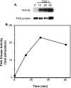
Activation of PAK by TSP-1 adhesion. C2C12 myoblastic cells were suspended for 60 min in BSA-blocked dishes (0 timepoint), or adhered to TSP-1 for times between 13 and 60 min. PAK activity was measured by in gel kinase assay and PAK protein level determined by Western blot (A). Stimulation of kinase activity relative to suspended cells was quantitated by image scanning and is presented normalized according to PAK protein levels (B). The data are representative of three independent experiments.
Figure 7
Figure 7
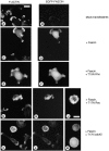
Effects of dominant-negative GTPases on fascin microspike formation by TSP-1-adherent C2C12 cells. Cells transiently transfected with pCMV expression vector (mock-transfectants; A and B), or expression vectors encoding EGFP-fascin alone (C and D), or T19N Rho plus EGFP-fascin (E and F), T17N Rac plus EGFP-fascin (G, H, and J) or T17N Cdc42 plus EGFP-fascin (K–M) were analyzed after 1 h adhesion to 50 nM TSP-1. Transfectants were identified by EGFP-fascin expression and actin organization was determined by TRITC-phalloidin staining. The results shown are representative of those obtained in three independent experiments. Bars: (A, B, G, H, K, and L) 10 μm; (C, D, E, F, J, and M) 5 μm.
Figure 8
Figure 8
Effects of Rac or Cdc42 constructs on fascin spike formation by TSP-1–adherent C2C12 cells. (A) Effects of dominant-negative or constitutively active GTPases on cell attachment to TSP-1 at 1 h. The graph shows the mean ± SEM of data from three independent experiments and compares the percentage of transfected cells which attached to TSP-1 with the percentage of transfectants in the starting populations. (B) Cytoskeletal organization in C2C12 cells overexpressing Q61L Cdc42 or Q61L Rac. Cells were stained with TRITC-phalloidin after 1 h of adhesion to TSP-1 and positive cells were identified by EGFP-fascin fluorescence. The results shown are representative of those obtained in three independent experiments. Bar, 5 μm.
Figure 8
Figure 8
Effects of Rac or Cdc42 constructs on fascin spike formation by TSP-1–adherent C2C12 cells. (A) Effects of dominant-negative or constitutively active GTPases on cell attachment to TSP-1 at 1 h. The graph shows the mean ± SEM of data from three independent experiments and compares the percentage of transfected cells which attached to TSP-1 with the percentage of transfectants in the starting populations. (B) Cytoskeletal organization in C2C12 cells overexpressing Q61L Cdc42 or Q61L Rac. Cells were stained with TRITC-phalloidin after 1 h of adhesion to TSP-1 and positive cells were identified by EGFP-fascin fluorescence. The results shown are representative of those obtained in three independent experiments. Bar, 5 μm.
Figure 9
Figure 9
Effects of pharmacological inhibition of actin turnover on cell-matrix spreading and fascin spike formation. C2C12 cells were stained with TRITC-phalloidin after 1 h adhesion to TSP-1 (A and B) or fibronectin (C–F). Control cells (A and C) were compared with cells pretreated with 0.5 μm latrunculin B for 60 min (B and D) or 0.5 μM jasplakinolide for 30 min (E). Bar, 5 μm.
Figure 10
Figure 10
Characterization of anti-fascin immunoglobulins. (A) Schematic diagram of the fascin molecule showing the two actin-binding domains (ABD1 and ABD2), the MARCKS homology motif (MHM) and the locations of the synthetic peptides used to produce fascin polyclonal immunoglobulins. (B) Immunoprecipitation of fascin from metabolically labeled cell extracts by FAS-N and FAS-C immunoglobulins. Lane 1, mixed preimmune FAS-N and FAS-C immunoglobulins; lane 2, FAS-N IgG; lane 3, FAS-N preincubated with 10 ug of FAS-N peptide; lane 4, FAS-C IgG; lane 5, FAS-C IgG preincubated with 10 μg of FAS-C peptide; lane 6, in vitro translated fascin. (C) Effect of the antibodies on the binding of fascin to actin in a blot-overlay assay. Lane 1, 15 μg actin stained with Coomassie blue; lanes 2–8, autoradiographic detection of fascin bound to actin on nitrocellulose membrane. 5 μg of actin was loaded per lane. Overlay conditions were fascin only (lane 2), fascin plus mixed preimmune IgGs (lane 3), fascin plus mixed preimmune IgGs plus 10 μg each FAS-N and FAS-C peptides (lane 4), fascin plus FAS-N IgG (lane 5), fascin plus FAS-C IgG (lane 6), fascin plus FAS-N and FAS-C IgGs (lane 7) and fascin plus FAS-N and FAS-C IgGs plus 10 μg each of FAS-N and FAS-C peptides (lane 8).
Figure 11
Figure 11
Role of fascin organization in cell adhesion and motility. (A–K) C2C12 cells were loaded with 0.5 mg/ml nonimmune rabbit IgG (A and F); 0.5 mg/ml FAS-N IgG (B and G); 0.5 mg/ml FAS-C IgG (C and H); a 1:1 mixture of FAS-N and FAS-C at 0.5 mg/ml total (D and J), or a 1:1 mixture of 0.5 mg/ml FAS-N and FAS-C containing 50 ug/ml each FAS-N and FAS-C peptides (E and K). Cells were stained with TRITC-phalloidin after 1 h adhesion on 50 nM TSP-1 (A–E) or 50 nM FN (F–K). Bar, 10 um. (L and M) Quantitation of cell attachment at 1 h on 50 nM TSP-1 (L) or 50 nM FN (M). (L–P) Lane 1, cells loaded with 0.5 mg/ml nonimmune rabbit IgG; lane 2, cells loaded with 0.5 mg/ml FAS-N IgG; lane 3, cells loaded with 0.5 mg/ml FAS-C IgG (C and G); lane 4, cells loaded with a 1:1 mixture of FAS-N and FAS-C 0.5 mg/ml total; lane 5, cells loaded with a 1:1 mixture of FAS-N plus FAS-C at 0.2 mg/ml total; lane 6, cells loaded with a 1:1 mixture of 0.5 mg/ml FAS-N and FAS-C plus 14 μg/ml each of FAS-N and FAS-C peptides. Values shown are mean ± SEM from triplicate determinations. N, Effects of fascin antibodies on random C2C12 movement on a 50 nM TSP-1 substratum. P, Effects of fascin antibodies on random C2C12 cell movement on a 50-nM FN substratum. Values are mean ± SEM from duplicate experiments. An asterisk indicates mean value significantly different from control sample at P = 0.001.
Figure 11
Figure 11
Role of fascin organization in cell adhesion and motility. (A–K) C2C12 cells were loaded with 0.5 mg/ml nonimmune rabbit IgG (A and F); 0.5 mg/ml FAS-N IgG (B and G); 0.5 mg/ml FAS-C IgG (C and H); a 1:1 mixture of FAS-N and FAS-C at 0.5 mg/ml total (D and J), or a 1:1 mixture of 0.5 mg/ml FAS-N and FAS-C containing 50 ug/ml each FAS-N and FAS-C peptides (E and K). Cells were stained with TRITC-phalloidin after 1 h adhesion on 50 nM TSP-1 (A–E) or 50 nM FN (F–K). Bar, 10 um. (L and M) Quantitation of cell attachment at 1 h on 50 nM TSP-1 (L) or 50 nM FN (M). (L–P) Lane 1, cells loaded with 0.5 mg/ml nonimmune rabbit IgG; lane 2, cells loaded with 0.5 mg/ml FAS-N IgG; lane 3, cells loaded with 0.5 mg/ml FAS-C IgG (C and G); lane 4, cells loaded with a 1:1 mixture of FAS-N and FAS-C 0.5 mg/ml total; lane 5, cells loaded with a 1:1 mixture of FAS-N plus FAS-C at 0.2 mg/ml total; lane 6, cells loaded with a 1:1 mixture of 0.5 mg/ml FAS-N and FAS-C plus 14 μg/ml each of FAS-N and FAS-C peptides. Values shown are mean ± SEM from triplicate determinations. N, Effects of fascin antibodies on random C2C12 movement on a 50 nM TSP-1 substratum. P, Effects of fascin antibodies on random C2C12 cell movement on a 50-nM FN substratum. Values are mean ± SEM from duplicate experiments. An asterisk indicates mean value significantly different from control sample at P = 0.001.

Comment in

References

    1. Adams J.C. Formation of stable microspikes containing actin and the 55kDa actin-bundling protein, fascin, is a consequence of cell adhesion to thrombospondin-1implications for the antiadhesive activities of thrombospondin-1. J. Cell Sci. 1995;108:1977–1990. - PubMed
    1. Adams J.C. Characterisation of cell-matrix adhesion requirements for the formation of fascin microspikes. Mol. Biol. Cell. 1997;8:2345–2363. - PMC - PubMed
    1. Adams J.C., Tucker R.P., Lawler J. The thrombospondin gene family 1995. R.G. Landes; Austin, TX: pp. 188
    1. Adams J.C., Clelland J.D., Collett G.D.M., Matsumura F., Yamashiro S., Zhang L. Cell-matrix adhesions differentially regulate fascin phosphorylation. Mol. Biol. Cell. 1999;10:4177–4190. - PMC - PubMed
    1. Arber S., Barbayannis F.A., Hanser H., Schneider C., Stanyon C.A., Bernard O., Caroni P. Regulation of actin dynamics through phosphorylation of cofilin by LIM-kinase. Nature. 1998;393:805–809. - PubMed

Publication types

MeSH terms