. 2000 Aug 21;150(4):887-94.
doi: 10.1083/jcb.150.4.887.

Cross-talk between two cysteine protease families. Activation of caspase-12 by calpain in apoptosis

Affiliations

Cross-talk between two cysteine protease families. Activation of caspase-12 by calpain in apoptosis

T Nakagawa et al. J Cell Biol. .

Abstract

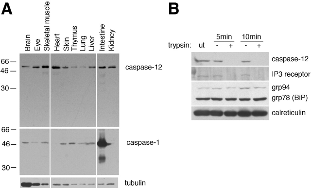
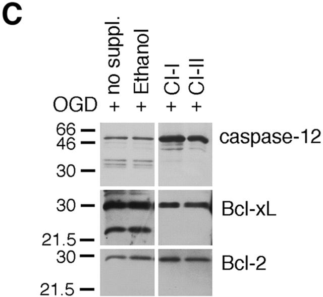
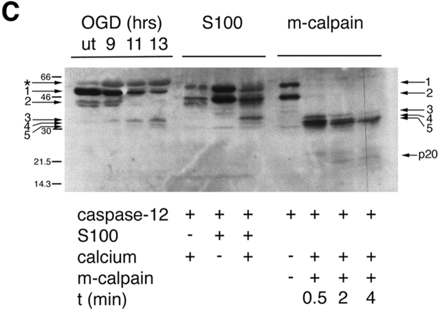
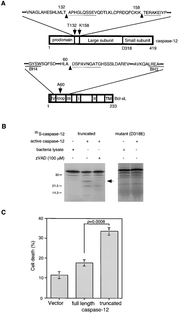
Calpains and caspases are two cysteine protease families that play important roles in regulating pathological cell death. Here, we report that m-calpain may be responsible for cleaving procaspase-12, a caspase localized in the ER, to generate active caspase-12. In addition, calpain may be responsible for cleaving the loop region in Bcl-xL and, therefore, turning an antiapoptotic molecule into a proapoptotic molecule. We propose that disturbance to intracellular calcium storage as a result of ischemic injury or amyloid beta peptide cytotoxicity may induce apoptosis through calpain- mediated caspase-12 activation and Bcl-xL inactivation. These data suggest a novel apoptotic pathway involving calcium-mediated calpain activation and cross-talks between calpain and caspase families.


Figures

Figure 1
Figure 1
The expression of caspase-12. (A) A Western blot of caspase-12. Anti–caspase-1 and antitubulin antibodies were used as controls. Caspase-12 is constitutively expressed in postnatal day 4 mouse tissues. (B) Caspase-12 localizes on the outer membrane of the ER. IP3R, a transmembrane protein, was readily digested by trypsin as that of caspase-12, whereas ER lumenal proteins, grp78, grp94, and calreticulin were protected against trypsin digestion. ut, untreated.
Figure 2
Figure 2
The requirement of calpain for caspase-12 activation in vivo. (A) Caspase-12 and Bcl-xL are cleaved in glial cells under oxygen and glucose deprivation (OGD, 9-h treatment), but not under hypoxia condition (oxygen deprivation, OD). ER stress is induced by OGD but not by OD, as indicated by the induction of BiP in OGD but not OD-treated cells. Antitubulin was used as a control (bottom). UT; untreated. (B) Caspase-12 is cleaved in glial cells treated by ER stress–inducing agents (thapsigargin, 2 μM; A23187, 2 μM), which induces ER stress as indicated by BiP induction. (C and D) The effects of pan-caspase inhibitor (zVAD), and calpain inhibitor (calpain inhibitors I, II, and E64d) in glial cells treated by OGD. Calpain inhibitors (calpain inhibitors I and II; CI-I and -II, respectively), but not z-VAD, inhibited caspase-12 and Bcl-xL cleavages (C and D). zVAD and calpain inhibitor partially protected glial cell death induced by OGD (D). Calpain activation correlates with caspase-12 cleavage activity. Endogenous calpain substrate, α-spectrin, was cleaved to 145- and 150-kD fragments after OGD treatment (D), which indicates calpain activation. (E) Fibrillar Aβ peptide induced caspase-12 cleavage, and calpain inhibitors prevented caspase-12 cleavage in primary cortical neurons. The cleavage product of caspase-12 was detected by Western blotting after 48 h of treatment with 40 μM fibrillar Aβ (25-35). Arrows indicate cleaved caspase-12 fragment. no suppl: no supplement.
Figure 2
Figure 2
The requirement of calpain for caspase-12 activation in vivo. (A) Caspase-12 and Bcl-xL are cleaved in glial cells under oxygen and glucose deprivation (OGD, 9-h treatment), but not under hypoxia condition (oxygen deprivation, OD). ER stress is induced by OGD but not by OD, as indicated by the induction of BiP in OGD but not OD-treated cells. Antitubulin was used as a control (bottom). UT; untreated. (B) Caspase-12 is cleaved in glial cells treated by ER stress–inducing agents (thapsigargin, 2 μM; A23187, 2 μM), which induces ER stress as indicated by BiP induction. (C and D) The effects of pan-caspase inhibitor (zVAD), and calpain inhibitor (calpain inhibitors I, II, and E64d) in glial cells treated by OGD. Calpain inhibitors (calpain inhibitors I and II; CI-I and -II, respectively), but not z-VAD, inhibited caspase-12 and Bcl-xL cleavages (C and D). zVAD and calpain inhibitor partially protected glial cell death induced by OGD (D). Calpain activation correlates with caspase-12 cleavage activity. Endogenous calpain substrate, α-spectrin, was cleaved to 145- and 150-kD fragments after OGD treatment (D), which indicates calpain activation. (E) Fibrillar Aβ peptide induced caspase-12 cleavage, and calpain inhibitors prevented caspase-12 cleavage in primary cortical neurons. The cleavage product of caspase-12 was detected by Western blotting after 48 h of treatment with 40 μM fibrillar Aβ (25-35). Arrows indicate cleaved caspase-12 fragment. no suppl: no supplement.
Figure 3
Figure 3
Cleavage of caspase-12 by calpain in vitro. (A) Cleavage of caspase-12 by cerebral cortex–soluble (S-100) proteins in a calcium-dependent manner. Caspase-12 is effectively cleaved in the presence of millimolar, but not micromolar, calcium. Activation of caspase-12 is inhibited by calcium chelators (EGTA and EDTA). (B) Caspase-12 and Bcl-xL are cleaved by purified m-calpain in vitro. (a) Caspase-12 is cleaved by m-calpain to generate three major fragments each ∼35 kD, which may autoactivate themselves to generate fragments of 20 and 10 kD (arrows), which may be the large and small subunits of caspase-12, respectively. The 20-kD fragment is detected by anti–caspase-12 p20 antibody after a longer incubation with calpain (b). (c) Bcl-xL is cleaved by m-calpain in vitro. The arrow indicates the cleaved product that was sequenced. (C) The cleavage products of caspase-12 in vivo are similar in sizes to those generated by in vitro cleavage assays (S100 and purified m-calpain). The arrows point to the procaspase-12 (arrows 1 and 2) and the cleaved products of caspase-12 (arrows 3–5). Cleaved products (arrows 3 and 5) were sequenced. Cleavage sites of 3 and 5 are T132/A133 and K158/T159, respectively. The large subunit of caspase-12 (p20) is produced in a time-dependent manner.
Figure 3
Figure 3
Cleavage of caspase-12 by calpain in vitro. (A) Cleavage of caspase-12 by cerebral cortex–soluble (S-100) proteins in a calcium-dependent manner. Caspase-12 is effectively cleaved in the presence of millimolar, but not micromolar, calcium. Activation of caspase-12 is inhibited by calcium chelators (EGTA and EDTA). (B) Caspase-12 and Bcl-xL are cleaved by purified m-calpain in vitro. (a) Caspase-12 is cleaved by m-calpain to generate three major fragments each ∼35 kD, which may autoactivate themselves to generate fragments of 20 and 10 kD (arrows), which may be the large and small subunits of caspase-12, respectively. The 20-kD fragment is detected by anti–caspase-12 p20 antibody after a longer incubation with calpain (b). (c) Bcl-xL is cleaved by m-calpain in vitro. The arrow indicates the cleaved product that was sequenced. (C) The cleavage products of caspase-12 in vivo are similar in sizes to those generated by in vitro cleavage assays (S100 and purified m-calpain). The arrows point to the procaspase-12 (arrows 1 and 2) and the cleaved products of caspase-12 (arrows 3–5). Cleaved products (arrows 3 and 5) were sequenced. Cleavage sites of 3 and 5 are T132/A133 and K158/T159, respectively. The large subunit of caspase-12 (p20) is produced in a time-dependent manner.
Figure 4
Figure 4
The cleavage sites in caspase-12 and Bcl-xL by m-calpain. (A) Caspase-12 is cleaved in at least two sites at T132/A133 and K158/T159. Bcl-xL is cleaved at one site, A60/D61. The cleavage site in Bcl-xL is close to that of caspase-1 or -3, (D61/S62 by caspase-1 and -3, D76/A77 by caspase-3; Clem et al. 1998; Fujita et al. 1998). The cleavage site of Bcl-xL is not conserved in Bcl-2. Underlines show sequence results. (B) Truncated (T159-N419) caspase-12 (t-caspase-12) expressed in bacteria is active in cleaving the wild-type caspase-12 but not D318E mutant. Caspase-12 activity is inhibited by zVAD. Arrow indicates the cleaved product of caspase-12. (C) t-Caspase-12 induced cell death in Rat1 cells. The percentage of cell death was determined by morphology 24 h after transfection. Data are averages (± SEM).

References

    1. Alzheimer's Disease Collaborative Group The structure of the presenilin 1 (S182) gene and identification of six novel mutations in early onset AD families. Nat. Genet. 1995;11:219–222. - PubMed
    1. Begley J.G., Duan W., Chan S., Duff K., Mattson M.P. Altered calcium homeostasis and mitochondrial dysfunction in cortical synaptic compartments of presenilin-1 mutant mice. J. Neurochem. 1999;72:1030–1039. - PubMed
    1. Berridge M.J. Inositol trisphosphate and calcium signalling. Nature. 1993;361:315–325. - PubMed
    1. Berridge M.J., Bootman M.D., Lipp P. Calciuma life and death signal. Nature. 1998;395:645–648. - PubMed
    1. Bialik S., Cryns V.L., Drincic A., Miyata S., Wollowick A.L., Srinivasan A., Kitsis R.N. The mitochondrial apoptotic pathway is activated by serum and glucose deprivation in cardiac myocytes. Circ. Res. 1999;85:403–414. - PubMed

Publication types

MeSH terms