. 2000 Aug;106(4):501-9.
doi: 10.1172/JCI9148.

Leptin enhances wound re-epithelialization and constitutes a direct function of leptin in skin repair

Affiliations

Leptin enhances wound re-epithelialization and constitutes a direct function of leptin in skin repair

S Frank et al. J Clin Invest. 2000 Aug.

Abstract

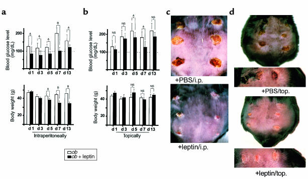
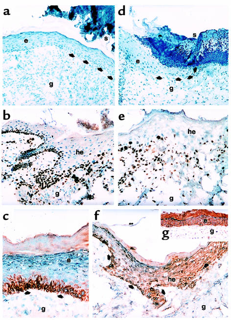
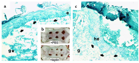
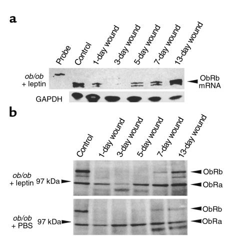
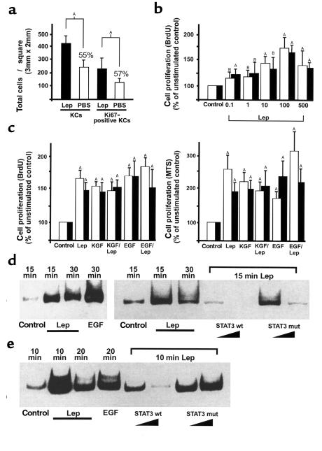
Wound-healing disorders are a therapeutic problem of extensive clinical importance. Leptin-deficient ob/ob mice are characterized by a severely delayed wound healing that has been explained by the mild diabetic phenotype of these animals. Here we demonstrate that systemically and topically supplemented leptin improved re-epithelialization of wounds in ob/ob mice. Leptin completely reversed the atrophied morphology of the migrating epithelial tongue observed at the wound margins of leptin-deficient animals into a well-organized hyperproliferative epithelium. Moreover, topically supplemented leptin accelerated normal wound-healing conditions in wild-type mice. As assessed by immunohistochemistry, proliferating keratinocytes located at the wound margins specifically expressed the leptin-receptor subtype ObRb during repair. Additionally, leptin mediated a mitogenic stimulus to the human keratinocyte cell line HaCaT and human primary keratinocytes in vitro. Therefore, leptin might represent an effective novel therapeutic factor to improve impaired wound-healing conditions.


Figures

Figure 1
Figure 1
Effect of intraperitoneally and topically supplemented leptin on blood glucose level, body weight, and wound healing in ob/ob mice. C57BL/6J-ob/ob mice were treated with leptin intraperitoneally (i.p.) or topically (top) as described in Methods. PBS-treated ob/ob mice were used as a control. (a and b) Blood glucose levels and body weight were determined for intraperitoneally injected (a) or topically treated (b) animals at the indicated experimental time points. Three animals (n = 3) were analyzed at every experimental time point. Data are expressed as milligrams per deciliter (for blood glucose levels) or grams (for body weight). Mean changes ± SD in blood glucose levels, or body weight, respectively, are shown. AP < 0.05; BP < 0.01; NS, not significant compared with the conditions as indicated with the brackets. (c) Thirteen-day wounds after intraperitoneal (i.p.) treatment of animals with 5 μg leptin/g of body weight (+leptin/i.p.) or PBS (+PBS/i.p.). (d) Back skin of representative C57BL/6J-ob/ob mice topically treated with 1 μg leptin in 20 μL PBS twice a day (+leptin/top.) or PBS (+PBS/top.) at day 10 after wounding. The bottom panels demonstrate the same wounds in higher magnification.
Figure 2
Figure 2
Effect of leptin on epithelial proliferation and localization of leptin-receptor subtypes in the skin. (a and d) Hematoxylin/eosin-stained frozen sections from 10-day wounds isolated from the same C57BL/6J-ob/ob individual that had been treated topically with 1 μg leptin/20 μL PBS twice a day on the left-side wounds (a) or PBS only on the right-side wounds (d), respectively. Arrows indicate the leading edge of the migrating epithelium. (b and e) Frozen sections from 5-day wounds isolated from leptin-treated (intraperitoneally) (b) or PBS-injected (e) C57BL/6J-ob/ob mice were incubated with a monospecific, polyclonal Ab directed against murine Ki67. (c) Frozen section from a 13-day wound isolated from a leptin-treated (intraperitoneally) mouse. The section was incubated with a monospecific polyclonal Ab against the ObRb-receptor subtype. (f) Frozen section from a 7-day wound isolated from a PBS-treated (intraperitoneally) mouse. The section was incubated with a monospecific polyclonal Ab against the ObRb-receptor subtype. Note that the epithelium did not extend into the granulation tissue (compare to b, epithelium of leptin-treated mouse, 5 days). (g) Frozen section from a 13-day wound isolated from a wild-type mouse (BALB/c). The section was incubated with a monospecific polyclonal Ab recognizing the ObRb- and ObRa-receptor subtypes. Sections were stained with the avidin-biotin-peroxidase complex system using 3-amino-9-ethylcarbazole as a chromogenic substrate (b, c, e, f, g). Nuclei were counterstained with hematoxylin. Strongly immunopositive signals within the sections are indicated with arrows. e, epithelium; g, granulation tissue; he, hyperproliferative epithelium; s, scab.
Figure 3
Figure 3
Effect of topically supplemented leptin on wound healing in wild-type mice. Balb/c mice were topically treated with leptin as described in Methods. PBS-treated mice were used as a control. (b) Wounded back skin of representative Balb/c mice (5 days after wounding). Wounds have been treated topically for the whole 5-day period after wounding with 5 μg leptin/20 μL PBS twice a day (+leptin) or 20 μL PBS (+PBS), respectively. (a and c) Hematoxylin/eosin-stained frozen sections from 5-day wounds isolated from Balb/c mice that have been treated topically with 5 μg leptin/20 μL PBS twice a day (a) or 20 μL PBS only (c). Arrows indicate the leading edge of the migrating epithelium. e, epithelium; g, granulation tissue; gH, atrophied granulation tissue; he, hyperproliferative epithelium; s, scab.
Figure 4
Figure 4
Regulation of leptin-receptor expression during wound healing in Balb/c (wt) and C57BL/6J-ob/ob mice. (a) C57BL/6J mice were intraperitoneally treated with leptin as described in Methods. Total cellular RNA (20 μg) from nonwounded and wounded back skin of C57BL/6J-ob/ob mice treated with leptin as indicated was analyzed by RNase protection assay with an RNA hybridization probe complementary to ObRb and ObRa leptin–receptor splice variants. For every experimental time point, three wounds each from three animals (total: n = 9 wounds) were pooled for analysis. The time after injury is indicated for each lane. Control skin refers to nonwounded skin. Hybridization probe (1000 counts/min) was added to the lane labeled probe. Expression of GAPDH mRNA is shown as a loading control in the bottom panel. (b) Total protein (50 μg) from lysates of nonwounded and wounded back skin (day 1, 3, 5, 7, and 13 after injury, indicated for each lane) of C57BL/6J-ob/ob mice treated with leptin or PBS (used as control) as indicated were analyzed by immunoblotting for the presence of leptin-receptor subtype proteins ObRa and ObRb. Two wounds from the backs of three animals (n = 6) were excised for each experimental time point and used for protein isolation. Leptin-receptor subtypes ObRa and ObRb are indicated by arrowheads.
Figure 5
Figure 5
Effect of leptin on proliferation and STAT3 activation in HaCaT keratinocytes and human primary keratinocytes. (a) Low-magnification photographs (100×) of frozen serial sections were analyzed for total keratinocyte cell numbers or proliferating keratinocytes within the hyperproliferative epithelia from leptin-treated (intraperitoneally) ob/ob mice and PBS-treated (intraperitoneally) ob/ob mice (n = 3), as indicated. This was done by counting hematoxylin-stained keratinocyte nuclei or Ki67-immunostained keratinocyte nuclei in a defined area (3 × 2 mm). Data are expressed as total number of keratinocyte nuclei ± SD (n = 3). AP < 0.01 as indicated by the brackets, percentage of means compared with leptin-treated animals. (b) Dose-dependent effects of human recombinant leptin (0.1–500 ng/mL) on HaCaT (open bars) or primary (filled bars) keratinocyte proliferation. Proliferation was determined by BrdU incorporation. Data are expressed as percentage of unstimulated control. Mean percentage of change in proliferation ± SD are shown (values represent the mean of five assays with a readout of nine wells for each concentration; n = 45). AP < 0.01 compared with control; BP < 0.05 compared with control. (c) Effects of leptin (100 ng/mL), KGF (10 ng/mL), or EGF (10 ng/mL) on HaCaT (open bars) and primary (filled bars) keratinocyte proliferation. KGF and leptin or EGF and leptin were also given simultaneously, as indicated. Proliferation was determined by BrdU incorporation (left panel) or using the MTS reagent (right panel) as described in Methods. Data are expressed as percentage of unstimulated control. Mean percentage of change in proliferation ± SD are shown (values represent the mean of six assays with a readout of nine wells for each condition; n = 54). AP < 0.01 compared with control. (d and e) EMSA. HaCaT keratinocytes (d) and primary keratinocytes (e) were grown to confluence and rendered quiescent by a 24-hour incubation in serum-free medium. Cells were subsequently stimulated with leptin (100 ng/mL) or EGF (10 ng/mL) for the indicated time periods. Nuclear extracts of stimulated cells were isolated as described in Methods. Specificity of binding was confirmed by competition experiments using a 10–100-fold excess of unlabeled wild-type (STAT3 wt) or mutated STAT3 (STAT3 mut) consensus oligo as indicated. Lep, leptin; KC, keratinocyte.

References

    1. Zhang Y, et al. Positional cloning of the mouse obese gene and its human homologue. Nature. 1994;372:425–432. - PubMed
    1. Masuzaki H, et al. Human obese gene expression. Adipocyte-specific expression and regional differences in the adipose tissue. Diabetes. 1995;44:855–858. - PubMed
    1. Halaas JL, et al. Weight-reducing effects of the plasma protein encoded by the obese gene. Science. 1995;269:543–546. - PubMed
    1. Vaisse C, et al. Leptin activation of STAT3 in the hypothalamus of wild-type and ob/ob mice but not db/db mice. Nat Genet. 1996;14:95–97. - PubMed
    1. Ghilardi N, et al. Defective STAT signaling by the leptin receptor in diabetic mice. Proc Natl Acad Sci USA. 1996;93:6231–6235. - PMC - PubMed

Publication types

MeSH terms