. 2000 Aug;106(4):571-8.
doi: 10.1172/JCI9087.

Blood-derived angioblasts accelerate blood-flow restoration in diabetic mice

Affiliations

Blood-derived angioblasts accelerate blood-flow restoration in diabetic mice

G C Schatteman et al. J Clin Invest. 2000 Aug.

Abstract

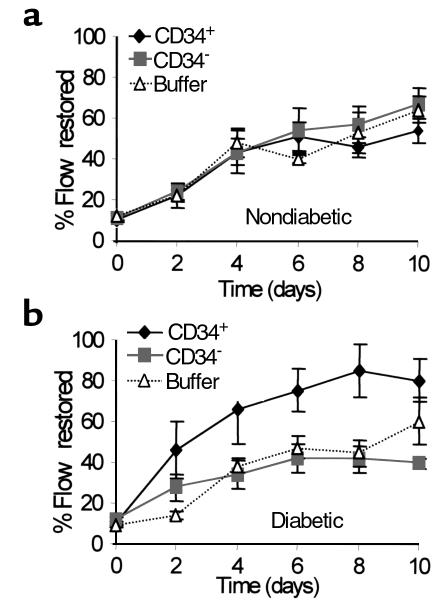
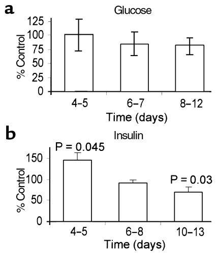
Endothelial cell progenitors, angioblasts, have been detected in the peripheral blood of adult humans, mice, and rabbits. These cells have been shown to incorporate into the endothelium of newly forming blood vessels in pathological and nonpathological conditions. Here we investigated the possibility that the CD34-expressing leukocytes (CD34(+) cells) that appear to be enriched for angioblasts could be used to accelerate the rate of blood-flow restoration in nondiabetic and diabetic mice undergoing neovascularization due to hindlimb ischemia. CD34(+) cells did not accelerate the restoration of flow in nondiabetic mice, but dramatically increased it in diabetic mice. Furthermore, CD34(+) cells derived from type 1 diabetics produced fewer differentiated endothelial cells in culture than did their type 2 diabetic- or nondiabetic-derived counterparts. In vitro experiments suggest that hyperglycemia per se does not alter the ability of angioblasts to differentiate or of angioblast-derived endothelial cells to proliferate. In contrast, hyperinsulinemia may enhance angioblast differentiation but impair angioblast-derived endothelial cell survival or proliferation. Our findings suggest that CD34(+) cells may be a useful tool for therapeutic angiogenesis in diabetics.


Figures

Figure 1
Figure 1
Laser Doppler blood-flow images. Representation of laser Doppler scans of diabetic mice after left hindlimb iliac artery ligation. Images are of diabetic mice injected with CD34+ cells (top) or CD34 cells (bottom) immediately after, 2 days after, and 8 days after surgery. The mice in the two series had fluxes near the mean for their respective groups at the 3 days shown. Red represents the highest flux and blue/gray the lowest.
Figure 2
Figure 2
Restoration of blood flow to surgically induced ischemic limbs. The data are expressed as the percentage of blood flow in the operated limb relative to the contralateral unoperated limb on the same day. Data were normalized to correct for preoperative differences in the two legs. The mean fluxes were determined using laser Doppler imaging for 7–10 mice in each group and were averaged. Error bars represent standard errors. (a) Nondiabetic mice. (b) Diabetic mice (serum glucose > 220 mg/dL). CD34+, injected with CD34+ cells; CD34, injected with CD34 cells; buffer, injected with vehicle or not injected.
Figure 3
Figure 3
CD34+ cells are present in ischemic limb vasculature. Confocal images of 10-μm sections of muscle from an ischemic limb of two diabetic mice treated with CM-DiI–labeled CD34+ cells. Sections from muscle collected 18 days after surgery were immunolabeled with FITC-conjugated anti–Tie-2 Ab. (a and b) The same section. (c and d) The same section. Confocal images of CM-DiI–labeled cells (white arrows) (a, c) that have formed vessels and are Tie-2 immunolabeled (black arrows) (b, d).
Figure 4
Figure 4
Anti-Tie-2 immunostaining of CD34+ cell cultures. Confocal microscope images of cells immunolabeled 14 days after plating on fibronectin with anti–Tie-2 (a) or rabbit IgG (b), and visualized with FITC.
Figure 5
Figure 5
Effects of glucose and insulin on cultured angioblasts. The number of spindle-shaped cells were counted in cultures of freshly isolated CD34+ cells plated on fibronectin at various times after plating. Results from four to six experiments at each time point, performed at least in duplicate, are pooled. (a) Cells cultured in medium or medium with 30 mM glucose. Data are presented as percentage of spindle-shaped cells in glucose-treated dishes relative to untreated controls. (b) Cells cultured in medium or medium with 6–12 μg/mL supplemental insulin. Data are presented as percentage of spindle-shaped cells in insulin-treated dishes relative to untreated controls. Error bars indicate standard errors for both a and b.

References

    1. Hueper WC, Russell MA. “Capillary-like formations” in tissue cultures of leukocytes. Archiv für Experimentelle Zellforschung Besoners Gewebezüchtung. 1932;12:407–424.
    1. Parker RC. The development of organized vessels in cultures of blood cells. Science. 1933;77:544–546. - PubMed
    1. White JF, Parshley MS. Growth in vitro of blood vessels from bone marrow of adult chickens. Am J Anat. 1950;89:321–345. - PubMed
    1. Hladovec J, Rossmann P. Circulating endothelial cells isolated together with platelets and the experimental modification of their counts in rats. Thromb Res. 1973;3:665–674.
    1. Solovey A, et al. Circulating activated endothelial cells in sickle cell anemia. N Engl J Med. 1997;337:1584–1590. - PubMed

Publication types

MeSH terms