

MR line-scan diffusion imaging of the spinal cord in children
Affiliations
- PMID: 10954293
- PMCID: PMC8174908
Item in Clipboard
MR line-scan diffusion imaging of the spinal cord in children
AJNR Am J Neuroradiol.
2000 Aug.
Abstract
Diffusion imaging has been widely used in the brain, but its application in the spinal cord has been limited. Using line-scan diffusion imaging (LSDI), a technique that is less sensitive to magnetic susceptibility and motion artifacts than are other diffusion techniques, we have successfully imaged the spinal cord in children. The apparent diffusion coefficient and relative diffusion anisotropy of the normal spinal cord were measured. LSDI was compared with echo-planar diffusion imaging of the spine in three patients.
Figures
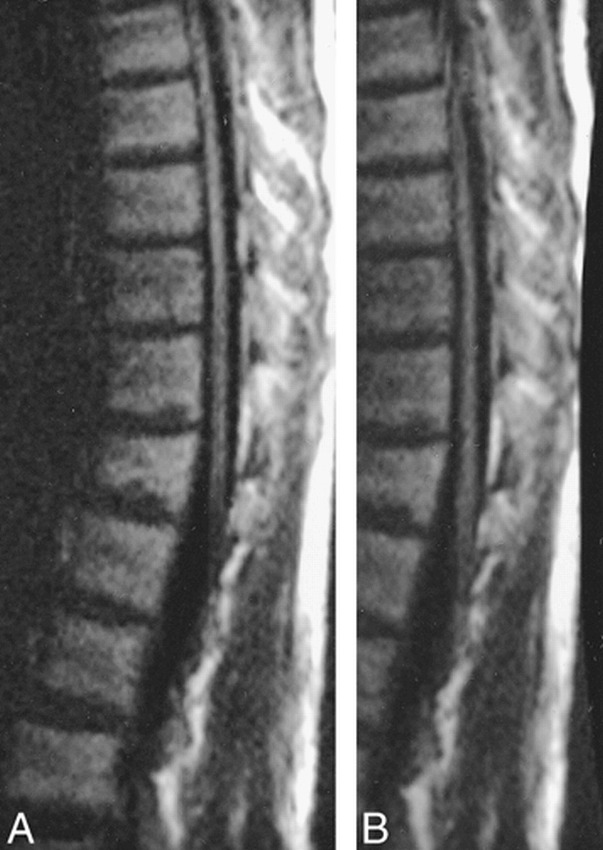
Comparison of LSDI images of the thoracic spine in a healthy adult volunteer shows comparable quality. A, Half field of view (64 columns). Sagittal LSDI isotropic high b factor image (2912/76/1, 128 × 64 columns, 4-mm section thickness, b = 750 s/mm2 extrapolated to 1000 s/mm2, six directions) using a 30 × 15-cm field of view and an imaging time of 50 s per location. B, Quarter field of view (32 columns). Sagittal LSDI isotropic high b factor image (1456/76/1, 128 × 32 columns, 4-mm section thickness, b = 750 s/mm2 extrapolated to 1000 s/mm2, six directions) using a 30 × 7.5-cm field of view and an imaging time of 25 s per location.
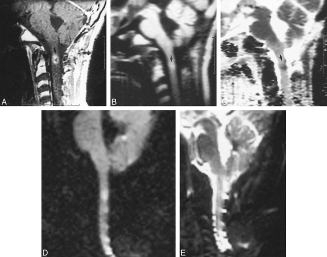
Case of a 16-year-old male patient with medulloblastoma treated with craniospinal radiation with presumed radiation effect at the site of overlap of the cranial and spinal radiation fields in the upper cervical spinal cord. Based on the intramedullary location of the lesion, metastatic medulloblastoma was considered unlikely. On follow-up images several months later (not shown) the lesion resolved. No anti-cancer therapy was undertaken during that time, and radiation effect was therefore considered to have been the most likely cause of the lesion. A, Sagittal contrast-enhanced T1-weighted image (600/20/2) of the cervical spine shows intramedullary enhancement (arrow). B, Sagittal LSDI isotropic high b factor diffusion image (2014/95/1, 4-mm section thickness, b = 750 s/mm2 extrapolated to 1000 s/mm2, six directions) shows increased signal within the lesion (arrow). C, Sagittal LSDI trace ADC map shows low intensity within the lesion confirming decreased diffusion (arrow). D, Sagittal EPDI isotropic high b factor diffusion image (4999/108/1, 5-mm section thickness, b =1000 s/mm2, three directions) shows artifactual high signal intensity in multiple locations in the cervical spinal cord. E, Sagittal EPDI trace ADC map is degraded by artifact and does not show the lesion seen on both the conventional MR and the LSDI sequences.
Comment in
-
Diffusion-weighted imaging of the spinal cord: is there a future?AJNR Am J Neuroradiol. 2000 Aug;21(7):1181-2. AJNR Am J Neuroradiol. 2000. PMID: 10954264 Free PMC article. No abstract available.
References
Publication types
MeSH terms
LinkOut - more resources
Full Text Sources
Medical